
Page 161 - Read Online
P. 161
Saneto. J Transl Genet Genom 2020;4:384-428 I http://dx.doi.org/10.20517/jtgg.2020.40 Page 389
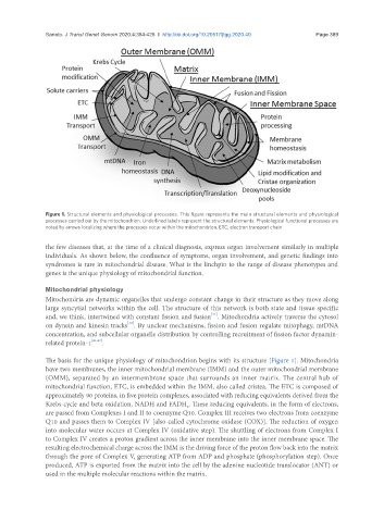
Figure 1. Structural elements and physiological processes. This figure represents the main structural elements and physiological
processes carried out by the mitochondrion. Underlined labels represent the structural elements. Physiological functional processes are
noted by arrows localizing where the processes occur within the mitochondrion. ETC: electron transport chain
the few diseases that, at the time of a clinical diagnosis, express organ involvement similarly in multiple
individuals. As shown below, the confluence of symptoms, organ involvement, and genetic findings into
syndromes is rare in mitochondrial disease. What is the linchpin to the range of disease phenotypes and
genes is the unique physiology of mitochondrial function.
Mitochondrial physiology
Mitochondria are dynamic organelles that undergo constant change in their structure as they move along
large syncytial networks within the cell. The structure of this network is both state and tissue specific
[41]
and, we think, intertwined with constant fission and fusion . Mitochondria actively traverse the cytosol
[42]
on dynein and kinesin tracks . By unclear mechanisms, fission and fusion regulate mitophagy, mtDNA
concentration, and subcellular organelle distribution by controlling recruitment of fission factor dynamin-
related protein-1 [41,43] .
The basis for the unique physiology of mitochondrion begins with its structure [Figure 1]. Mitochondria
have two membranes, the inner mitochondrial membrane (IMM) and the outer mitochondrial membrane
(OMM), separated by an intermembrane space that surrounds an inner matrix. The central hub of
mitochondrial function, ETC, is embedded within the IMM, also called cristea. The ETC is composed of
approximately 90 proteins, in five protein complexes, associated with reducing equivalents derived from the
Krebs cycle and beta oxidation, NADH and FADH . These reducing equivalents, in the form of electrons,
2
are passed from Complexes I and II to coenzyme Q10. Complex III receives two electrons from coenzyme
Q10 and passes them to Complex IV [also called cytochrome oxidase (COX)]. The reduction of oxygen
into molecular water occurs at Complex IV (oxidative step). The shuttling of electrons from Complex I
to Complex IV creates a proton gradient across the inner membrane into the inner membrane space. The
resulting electrochemical charge across the IMM is the driving force of the proton flow back into the matrix
through the pore of Complex V, generating ATP from ADP and phosphate (phosphorylation step). Once
produced, ATP is exported from the matrix into the cell by the adenine nucleotide translocator (ANT) or
used in the multiple molecular reactions within the matrix.