
Page 164 - Read Online
P. 164
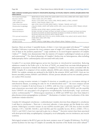
Page 392                                             Saneto. J Transl Genet Genom 2020;4:384-428  I  http://dx.doi.org/10.20517/jtgg.2020.40

               Table 4. Nuclear-encoded genes involved in mitochondrial physiology not directly related to oxidative phosphorylation that
               induce disease
                Fe-S cluster biosynthesis:  ABCB7, BOLA3, FDX1L, FDXR, FXN, GLRX5, IBA57, ISCA2, ISCU, LYRM4, NFS1, NFU1, IREB2, C19orf12
                Enzyme co-factors:    COASY, FLAD1, LIAS, LIPT1, PANK2
                Protein quality control:  AFG3L2, CLPP, LONP1, SPG7, YME1L1, PARL, PMPCB, IMMP2L, HTRA2, XPNPEP3
                Lipid modification:   AAD3A, CHKB, PLA2G6, SERAC1, TAZ
                Protein Import/processing:  AKG, AIFM1, DNAJC19, GFER, MIPEP, PMPCA, TIMM8A, TIMM50, AMT, GLDC
                Mitochondrial morphology:  C19orf70, DNM1L, GDAP1, MFF, MFN2, MSTO1, OPA1, SACS, SLC25A46, STAT2, TRANK1, VPS13D,
                                      VPS13A
                Matrix metabolism:    D2HGDH, ECHS1, ETHE1, HIBCH, L2HGDH, NAKE, TXN2
                Metabolic transport:  SLC19A1, SLC25A1, SLC25A3, SLC25A4, SLC25A10, SLC25A12, SLC25A13, SLC25A15, SLC25A16,
                                      SLC25A19, SLC25A20, SLC25A21, SLC25A22, SLC25A24, SLC25A26, SLC25A32, SLC25A38,
                                      SLC25A42, SLC25A46, SLC2A13, MICU1, MICU2, MPC1
                TCA cycle and metabolism:  ACO2, ALDH18A1, DLAT, DLD, FH, HAAO, IDH3A, IDH3B, KYNU, MDH2, MECR, NADK2, PDHA1,
                                      PDHB, PDHX, PDK3, PDP1, PPA2
                Autoptosis/autophagy:  HTRA2, VPS13C
                Unclear function:     APOPT1, C19orf12, C1QBP, FBXL4, OPA3, RTN4IP1, SFXN4, TMEM65, CYP2U1


               function. There are at least 15 assembly factors, of which 11 have been associated with disease [58-62] . Isolated
               Complex I deficiency represents the most common cause of single ETC-induced disease, accounting for
                                                  [56]
               30% of them in the pediatric population . Leigh syndrome is the most frequent expression of Complex
               I defects [63,64] . Studies have demonstrated that the majority of Complex I pathological variants are private
                              [65]
               and nonrecurring . The clinical phenotype of Complex I dysfunction are heterogeneous, often including
               leukodystrophy and/or cardiomyopathy and associated with early death.

               Complex II or succinate dehydrogenase serves two functions in mitochondrial metabolism. Reducing
               substances created in the Krebs cycle, in the form of FADH , are passed from succinate dehydrogenase to
                                                                  2
               ubiquinone as succinate is formed into fumarate. Complex II is located in the matrix associated with the
               IMM. Complex II is composed of two catalytic subunits, SDHA and a ferrous sulfate containing subunit,
               SDHB. The catalytic subunits are anchored to the IMM by the subunits, SDHC and SCHD. There are two
               known assembly proteins, SDHAF1 and SDHAF2. All four protein subunits and the two assembly proteins
               are nuclear DNA-encoded.

               Disease-causing recessive variants in Complex II structure or assembly are an uncommon etiology of
                                                                               [66]
               mitochondrial disease, accounting for only 2%-8% of mitochondrial cases . The most common subunit
                                                                                               [67]
               involved in Complex II-derived disease is in SDHA, with less than 35 patients reported . The two
               main phenotypes associated with Complex II-encoded genes, SDHA, SDHB, SDHD, and the assembly
               factor SDHAF1 are associated with progressive encephalopathy leukodystrophy, Leigh syndrome,
               and/or cardiomyopathy [68,69] . Heterozygous mutations in SDHB and SDHD have also been linked to
               pheochromocytoma-paraganglioma syndromes . Mechanism of isolated tissue localization of the latter
                                                        [70]
               syndromes remains unknown.

               Complex III (ubinquinol-cytochrome c oxidoreductase) transfers electrons from ubiquinol to cytochrome
               b and then to cytochrome c. There are 11 structural subunits, 2 heme groups, and the Rieske iron-sulfur
               protein. Pathological recessive variants in one mtDNA-encoded gene, MT-CYB; four nuclear encoded
               subunits, CYC1, UQCRB, UQCRC2, and UQCRQ; and five assembly factors, BCS1L, LYRM7, TTC19,
                                                                     [71]
               UQCC2, and UQCC3, have been reported to give rise to disease . Recessive variants have been associated
               with developmental delay, encephalopathy, hepatopathy, renal tubulopathy, exercise intolerance, and muscle
               weakness.


               Pathological variants in the BCS1L gene are the most common cause of Complex III deficiency. The BCS1L
                                                                                                       [72]
               protein facilitates the last step of Complex III assembly, the insertion of the Rieske iron-sulfur subunit .