
Page 9 - Read Online
P. 9
Zhang et al. J Transl Genet Genom 2024;8:302-11 https://dx.doi.org/10.20517/jtgg.2024.39 Page 306
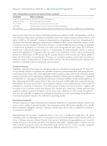
Table 2. Potential effects of enzymatic and supportive therapy on podocytes
Treatments Effects on podocytes
Enzyme replacement therapy A reduction in Gb3 levels within podocytes
Chaperone therapy Reduced Gb3 deposits on podocytes
SNCA inhibitor Improve lysosome podocyte structure and function
ACEI/ARB Decrease progressive kidney disease by alleviating podocyte injury
SGLT2 inhibitors Alleviate podocyte damage by targeting the pathogenetic mechanisms, such as oxidative stress and inflammation
Data from the Fabry Outcome Survey (FOS) indicated that early initiation of ERT with agalsidase α can slow
renal function deterioration and improve symptoms, and it also underscored the sustained efficacy and
safety of ERT in FD patients . Sustained administration of agalsidase β has been demonstrated to
[27]
effectively clear Gb3 from mesangial and glomerular endothelial cells . A dose-dependent clearance of Gb3
[28]
in podocytes was also reported . Research by Nowak et al. indicated that the licensed dosage of Agalsidase
[28]
[29]
β outperforms Agalsidase α in lowering Lyso-Gb3 levels among patients with classic FD . However,
Arends et al. confirmed the higher prevalence of neutralizing anti-drug antibodies (ADAs) in patients
treated with agalsidase β . Long-term ERT can result in the production of ADAs, which diminishes the
[30]
therapeutic effectiveness of ERT by altering the catalytic function of the enzyme and cellular uptake to
hasten the deterioration of renal function. Therefore, no conclusive evidence to show that one enzyme is
superior to another during nearly two decades of ERT treatment. The choice between β and α enzymes often
depends on individual responses and specific clinical contexts
Chaperone therapy
Migalastat, a pharmaceutical chaperone, has gained approval as the first oral medication for FD since 2016.
It is specifically indicated for patients with amenable mutations, predominantly those with attenuated, late-
onset forms of the disease who retain significant residual enzyme activity. Individuals with these genetic
variants rarely have severe renal disease, resulting in minimal or absent podocyte impairment . Migalastat
[31]
is a derivative of 1-deoxygalactonojirimycin and acts as a structural stabilizer for the terminal galactose of
Gb3, enhancing susceptible mutant forms of the α-Gal A enzyme. Additionally, this compound increases
and stabilizes the lysosomal activity , promoting the transport of susceptible mutant α-Gal A from the
[32]
endoplasmic reticulum to lysosomes. The enhancement of kidney and heart function, coupled with
increased α-Gal A enzyme activity and reduced Gb3 deposits after chaperone therapy, indicated that
migalastat could be a practical therapeutic choice and a secure substitute for ERT among FD patients .
[33]
Moreover, the capability of migalastat to penetrate the blood-brain barrier is promising, and its efficacy on
neurological symptoms remains to be confirmed in upcoming studies.
Developing treatments
Substrate reduction therapies function by decreasing the synthesis of accumulated substrate caused by the
lack of α-Gal A activity. A principal benefit of these treatments is their effectiveness regardless of the specific
genetic mutation causing the enzymatic deficiency. Notably, they can be used as a standalone treatment or
in conjunction with ERT .
[34]
Pegunigalsidase alfa represents a pegylated variant of the α-Gal A, produced through plant cell culture
techniques, and is recently approved in the European Union and United States for the treatment of FD. In
terms of efficacy outcomes, kidney biopsies from patients administered pegunigalsidase alfa showed a
marked decrease in Gb3 levels, and their renal function was stable . Multiple phase-3 clinical trials are
[35]
either actively in progress, such as the BALANCE study [NCT02795676], or have recently concluded,
including the BRIGHT study [NCT03180840] and the BRIDGE study [NCT03018730].