
Page 7 - Read Online
P. 7
Zhang et al. J Transl Genet Genom 2024;8:302-11 https://dx.doi.org/10.20517/jtgg.2024.39 Page 304
Table 1. Clinical manifestation of Fabry nephropathy
Laboratory Clinical manifestation
examination
Urinalysis Microalbuminuria in early phases, then progress to moderate to severe proteinuria in adulthood
Hematuria
Kidney function Glomerular hyperfiltration is an early marker, then a progressive decrease in GFR
Urine microscopy Mulberry cells with characteristic “Maltese cross bodies”, podocyturia
Tubular dysfunction α1-microglobulin, N-acetyl-β-glucosaminidase, and alanine aminopeptidase were elevated in early phases,
isosthenuria, distal renal tubular acidosis
Ultrasound findings Renal cysts, mainly parapelvic ones
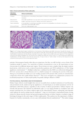
Figure 1. (A) The typical pathological manifestations of glomerulus in Fabry disease under light microscope. Vacuole-like changes in the
cytoplasm of podocytes and parietal epithelial cells were observed. (Periodic Acid-Schiff stain, ×400 times); (B) The typical pathological
manifestations of glomerulus in Fabry disease under light microscope. A large number of Gb3 bodies were deposited in the cytoplasm of
podocytes and parietal epithelial cells, and toluidine blue staining was observed. (×400 times); (C) Typical glomerular pathological
manifestations of Fabry disease under electron microscope. A large number of “zebra bodies” formed by Gb3 deposition in the
cytoplasm of podocytes and foot process segmental fusion.
patients. Heterozygous females often show no symptoms, but they can still develop a severe form of the
condition similar to males. The variability of female presentations is due to the inactivation of one
chromosome X in different tissues and organs. The processes are more complex than the simple random
inactivation, with factors such as skewed inactivation patterns and DNA methylation of GLA being
significant considerations [11,12] . Multiple studies have confirmed that the MSSI score effectively distinguishes
FD from various other critical illnesses and is a reliable measure for evaluating the severity of the pain.
Zhang et al. found that the MSSI score was notably elevated in FD patients with a decline in renal function
[13]
compared to those with stable kidney function . This score emerged as an independent predictor for
accelerated progression of Fabry nephropathy, frequently leading to dialysis or ESRD.
PATHOLOGICAL FEATURES AND PATHOGENESIS
Kidney biopsy offered crucial insights that were not accessible from standard evaluations of renal function
and proteinuria levels, underscoring the significance of renal biopsy as an initial assessment tool for all
individuals with Fabry disease, regardless of the severity of clinical presentation. Kidney biopsies can
identify and measure Gb3 deposits in individuals with a 30-300 mg/g albumin-to-creatinine ratio and
normal renal function. For women without signs of FD-related kidney disease, substantial renal deposits
could indicate the need to initiate specific treatment . Fogo et al. developed a standardized biopsy scoring
[14]
system for disease-specific lesions and general progression lesions in kidney biopsies, revealing a range of
histological changes in the early stages of Fabry nephropathy. This underscores the importance of kidney
[15]
biopsies for baseline assessments, even in cases with minimal clinical manifestations . The initial damage