
Page 160 - Read Online
P. 160
Page 4 of 7 Ioannides et al. J Cancer Metastasis Treat 2020;6:15 I http://dx.doi.org/10.20517/2394-4722.2020.22
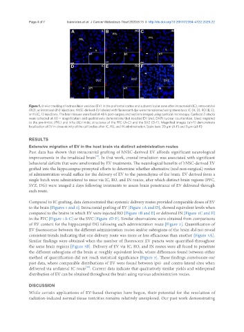
Figure 1. In vivo tracking of extracellular vesicles (EV) in the prefrontal cortex and subventricular zone after intracranial (IC), retro-orbital
(RO), or intranasal (IN) injections. hNSC-derived-EV labeled with fluorescent dye were transplanted using stereotaxic IC (A, D), RO (B, E),
or IN (C, F) injections. The brain tissues were fixed at 48 h post-surgery and sections imaged using confocal microscopy. Confocal Z-stacks
were collected at 60 × magnification and qualitatively demonstrate that injected EV (red, DAPI nuclear counterstain, blue) migrated
to the pre-limbic (PRL) and infra (IL) limbic structures of the PFC (A-C) and the SVZ (D-F). Magnified images (a1-f1) demonstrate
localization of EV in close vicinity of the cell bodies after IC, RO, and IN administration. Scale bars: 20 µm (A-F) and 3 µm (a1-f1)
RESULTS
Extensive migration of EV in the host brain via distinct administration routes
Past data has shown that intracranial grafting of hNSC-derived EV affords significant neurological
[9]
improvements in the irradiated brain . In that work, cranial irradiation was associated with significant
behavioral deficits that were ameliorated by EV treatments. The neurological benefits of hNSC-derived EV
grafted into the hippocampus prompted efforts to determine whether alternative (and non-surgical) routes
of administration would suffice for the delivery of EV to the parenchyma of the brain. EV derived from a
single batch were administered to mice via IC, RO, and IN routes, after which distinct brain regions (PFC,
SVZ, DG) were imaged 2 days following treatments to assess brain penetrance of EV delivered through
each route.
Compared to IC grafting, data demonstrated that systemic delivery routes provided comparable doses of EV
to the brain [Figures 1 and 2]. Intracranial grafting of EV [Figure 1A and D], showed equivalent levels when
compared to the brains in which EV were injected RO [Figure 1B and E] or delivered IN [Figure 1C and F]
in the PFC [Figure 1A-C] or the SVC [Figure 1D-F]. Similar observations were obtained from comparisons
of EV content for the hippocampal DG following each administration route [Figure 2]. Quantification of
EV fluorescence between the different administration routes and/or subregions of the brain did not reveal
consistent trends indicating that one delivery route was more or less efficacious than another [Figure 3A].
Similar findings were obtained when the number of fluorescent EV puncta were quantified throughout
the same brain regions [Figure 3B]. Delivery of EV via IC, RO, and IN routes were all found to penetrate
the different subregions of the brain at roughly equivalent levels, where differences found between either
method of quantification did not reach statistical significance [Figure 3]. These findings corroborate our
past data, where comparable distributions of EV were found between ipsi- and contra-lateral sites when
[19]
delivered via unilateral IC route . Current data indicate that qualitatively similar yields and widespread
distribution of EV can be obtained throughout the brain using various administration routes.
DISCUSSION
While certain applications of EV-based therapies have begun, their potential for the resolution of
radiation-induced normal tissue toxicities remains relatively unexplored. Our past work demonstrating