
Page 838 - Read Online
P. 838
Page 6 of 9 Wickremesekera et al. J Cancer Metastasis Treat 2019;5:62 I http://dx.doi.org/10.20517/2394-4722.2019.09
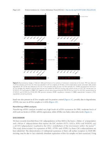
Figure 3. Representative images of Western blots performed on five samples of metastatic melanoma to the brain. PRR was detected
in all five samples at 40 kDa (A). ACE was detected in all five samples at 190 kDa, with multiple lower bands consistent with protein
degradation (B). ATIIR1 was detected at 45 kDa in two samples and at 28 kDa in all five samples (C). ATIIR2 was present at 50 kDa in
all five samples (H). Positive controls were mouse brain extract for PRR (A), mouse lung protein extract for ACE (B), mouse brain for
ATIIR1 (C), PC3 cell lysate for ATIIR2 (D). Negative controls were mouse kidney for PRR (A), human tonsil for ACE (B), mouse kidney for
ATIIR1 (C), and NTERA2 for ATIIR2 (D). PRR: pro-renin receptor; ATIIR1: angiotensin II receptor 1; ATIIR2: angiotensin II receptor 2; ACE:
angiotensin converting enzyme
band was also present in all five samples and the positive control [Figure 3C], possibly due to degradation.
ATIIR2 was seen in all five samples at 50 kDa [Figure 3D].
NanoString mRNA analysis
NanoString mRNA analysis revealed very high levels of mRNA expression for PRR, moderate levels of
ACE and low levels of ATIIR1 mRNA expression, while ATIIR2 was below detectable levels [Figure 4].
DISCUSSION
+
We have recently identified three CSC subpopulations within MM to the brain: a Melan-A subpopulation
-
and a Melan-A subpopulations that express the ESC markers OCT4, SALL4, SOX2 and NANOG, and
+
+
[21]
a pSTAT3 subpopulation localized to the CD34 endothelium of the microvessels within the tumor .
This study demonstrated the expression of PRR, ATIIR1 and ATIIR2 by these CSCs subpopulations we
have identified. The demonstration of widespread expression of these cell surface receptors by DAB IHC
staining, may be due to their relatively abundant expression within the samples we have examined. Using