
Page 836 - Read Online
P. 836
Page 4 of 9 Wickremesekera et al. J Cancer Metastasis Treat 2019;5:62 I http://dx.doi.org/10.20517/2394-4722.2019.09
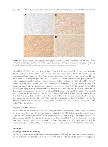
Figure 1. Representative DAB IHC-stained sections of metastatic melanoma to the brain showing endothelial staining of ACE (A,
brown), and mostly cytoplasmic staining of PRR (B, brown), ATIIR1 (C, brown) and ATIIR2 (D, brown). Nuclei were counter-stained with
hematoxylin (A-D, blue). Original magnification: 200×. DAB: 3, 3-diaminobenzidine; IHC: immunohistochemical; ATIIR1: angiotensin II
receptor 1; ATIIR2: angiotensin II receptor 2; PRR: pro-renin receptor; ACE: angiotensin converting enzyme
anti-ATIIR1 (AT2R1, 1:500; cat# sc-1173, Santa Cruz, CA, USA), anti-ATIIR2 (1:5000; cat# ab92445,
Abcam), anti-ACE (1:200; cat# sc-12184, Santa Cruz) and anti-β-actin (1:2000 cat# ab8226, Abcam).
Secondary antibodies used were Alexa Fluor 647 rabbit anti-mouse for β-actin (1:2000 cat# A21202, Thermo
TM
Fisher Scientific), donkey anti-goat HRP (1:10000; cat# ab97120; Abcam) and a rabbit anti-goat Superclonal
TM
biotin conjugated secondary antibody (1:20000; cat# A27013, Thermo Fisher Scientific) followed by a Pierce
Streptavidin Poly-HRP (1:5000, cat# 21140, Thermo Fisher Scientific) at 4 °C for 10 min for the ACE
TM
tertiary cascade. β-actin antibody probing was performed with the iBind Flex device (cat# SLF2000, Life
Technologies) using primary mouse monoclonal anti-β-actin (1:2000; cat# ab8226, Abcam) and secondary
donkey anti-mouse AlexaFluor 488 (1:2000; cat# A21202, Thermo Fisher Scientific). Clarity Western ECL
(cat# 1705061, Bio-Rad) was used to visualize HRP detected bands and the Chemi Doc MP Imaging System
(Bio-Rad) and Image Lab 5.0 software (Bio-Rad) were used for detection and analysis. Positive controls
were mouse brain extract for PRR and ATIIR1, mouse lung protein extract for ACE and PC3 cell lysate for
ATIIR2. Negative controls were mouse kidney for PRR, human tonsil for ACE, mouse kidney for ATIIR1
and NTERA2 for ATIIR2.
NanoString mRNA analysis
[27]
RNA extraction, as previously described , was performed on four snap-frozen samples of MM to
the brain from the same cohort of ten patients included in DAB IHC staining underwent. RNA was
TM
subjected to NanoString nCounter Gene Expression Assay (NanoString Technologies, Seattle, WA,
USA) as completed by New Zealand Genomics Ltd (Dunedin, NZ). Probes for the genes encoding
PRR (NM_005765.2), ATIIR1 (NM_000685.3), ATIIR2 (NM_000686.3), ACE (NM_000789.2) and the
housekeeping gene GAPDH (NM_002046.3) were designed and synthesized by NanoString Technologies.
RESULTS
Histology and DAB IHC staining
H&E staining (data not shown) demonstrated the presence of MM in all ten samples that stained positively
for the melanoma marker Melan-A (data not shown). The endothelium of the microvessels expressed