
Page 791 - Read Online
P. 791
Page 6 of 9 Zhang et al. J Cancer Metastasis Treat 2019;5:56 I http://dx.doi.org/10.20517/2394-4722.2018.112
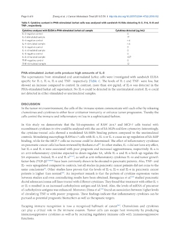
Table 1. Cytokine content in PHA-stimulated Jurkat cells was analyzed with sandwich ELISAs detecting IL-2, Il-6, IL-8 and
TNF-, respectively
Cytokines analyzed with ELISA in PHA-stimulated Jurkat cell sample Cytokines detected (pg/mL)
IL-2 negative control 0
IL-2 stimulated sample 17
IL-4 negative control n.d.
IL-4 stimulated sample n.d.
IL-6 negative control 0
IL-6 stimulated sample 0
IL-8 negative control 0
IL-8 stimulated sample 427
TNF-negative control 0
TNF-stimulated sample 12
PHA-stimulated Jurkat cells produce high amounts of IL-8
The supernatants from stimulated and unstimulated Jurkat cells were investigated with sandwich ELISA
specific for IL-2, IL-6, IL-8 and TNF- respectively [Table 1]. The levels of IL-2 and TNF- were low, but
showed an increase compared to control. In contrast, more than 400 pg/mL of IL-8 was detected in the
PHA-stimulated Jurkat cell supernatant. No IL-8 could be detected in the unstimulated control. IL-6 could
not detected in either stimulated or unstimulated samples.
DISCUSSION
In the tumor microenvironment, the cells of the immune system communicate with each other by releasing
chemokines and cytokines to either favor antitumor immunity or enhance tumor progression. Thereby the
cells control the immune and inflammatory milieu in a sophisticated fashion.
In this study we demonstrate that the SA-expression of RAW 264.7 and MCF-7 cells treated with
recombinant cytokines in vitro could be analyzed with the use of SA-MIPs and flow cytometry. Interestingly,
the cytokine-treated cells showed a modulated SA-MIPs binding pattern compared to the unstimulated
controls. Stimulating macrophage RAW264.7 cells with IL-4, IL-6 or IL-8 cause an up-regulation of SA-MIP
binding, while for the MCF-7 cells no increase could be determined. The effect of inflammatory cytokines
on pancreatic cancer cells has been reviewed by Roshani et al . In other studies, IL-4 did not have any effect,
[1]
but IL-6 and IL-8 were associated with poor prognosis and increased aggressiveness, respectively. IL-4 is
an anti-inflammatory cytokine expected to down-regulate SA, while IL-6 and IL-8 both up-regulate the
SA expression. Indeed, IL-6 and IL-8 [15-17] , as well as anti-inflammatory cytokines IL-10 and tumor growth
factor-beta (TGF-b) [15,18,19] have been commonly shown to be elevated in pancreatic patients. Also, TNF- and
IL- were upregulated compared to controls, but all studies in pancreatic cancer patients did not come to the
same conclusions . Other studies have proven that the levels of IL-4, IL-6 and IL-8 in pancreatic cancer
[1]
patients is higher than normal . An important remark is that the pattern of cytokine expression varies
[15]
between studies and even contradicting results have been obtained. Bassaganas et al. studied pancreatic
[20]
ductal adenocarcinoma cell lines treated with different cytokines. They found that treatment with either IL-1
or IL-6 resulted in an increased carbohydrate antigen and SA level. Also, the levels of mRNA of precursor
of carbohydrate antigens was enhanced. Moreover, Dima et al. found an association between higher levels
[19]
of circulating TNF-a with poorer prognosis. These findings indicate that inflammatory cytokines can be
pursued as potential prognostic biomarkers as well as therapeutic targets.
Escaping immune recognition is now a recognized hallmark of cancer . Chemokines and cytokines
[21]
can play a critical role in the immune evasion. Tumor cells can escape host immunity by producing
immunosuppressive cytokines as well as by recruiting regulatory immune cells with immunosuppressive
functions.