
Page 327 - Read Online
P. 327
Monks et al. J Cancer Metastasis Treat 2019;5:24 I http://dx.doi.org/10.20517/2394-4722.2018.79 Page 13 of 23
κ
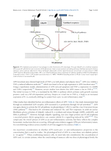
Figure 4. APN-mediated polarisation of macrophages to anti-inflammatory M2 phenotype. Through AdipoR1 and undefined receptors
APN causes NF-κB inhibition and promotion of M2 phenotype. Green arrows represent activating pathways. Red lines represent inhibitory
pathways. gAcrp: globular adiponectin; flAcrp: full length adiponectin; STAT6: signal transducer and activator of transcription 6; CREB:
cAMP response element-binding protein; JNK: c-Jun N-terminal kinase; SOC3: suppressor of cytokine signalling 3; Bcl3: B-cell lymphoma
3-encoded protein; TRAF1: TNF receptor-associated factor 1; TNIP3: TNFAIP3-interacting protein 3; NF-κB: nuclear factor kappa-light-
chain-enhancer of activated B cells
[5]
APN-knockout mice showed high levels of TNF-α in both plasma and adipose tissue . APN also inhibits a
TNF-α induced adhesion molecule [183] which was found to be in high expression in APN-knockout mice [184] .
Using a reperfusion model, administration of APN reduced apoptosis and TNF-α expression via AMPK
[18]
and COX-2 respectively . However, certain studies have shown that APN causes a rise in TNF-α [181,185] .
The initial increase in TNF-α is mediated by the ERK1/2 pathway which activates early-growth response
protein-1 and via a NF-κB-dependent pathway. Despite an initial rise in TNF-α, it leads to an increased
expression of IL-10, eventually shifting the system from a pro- to anti-inflammatory state [185] .
Other studies have identified further pro-inflammatory effects of APN [Table 2]. One study demonstrated that
through an unidentified APN receptor, APN increased IL-6 production through NF-κB activation . APN
[158]
was again shown to activate the NF-κB pathway via phospholipase C (PLC)-y and the c-Jun N-terminal Kinase
(JNK) pathway [186] . This lead to DC activation and enhanced Th1 and Th17 responses. In adult rat cardiac
fibroblasts, gAcrp activation of AdipoR1 induced IL-6 synthesis and release through AMPK, p38MAPK, and
[163]
ERK1/2 pathways . However, through AdipoR1 signalling, A20 (zinc finger protein) and B-cell lymphoma
3-encoded protein (Bcl3) upregulation can counter-inhibit IL-6 signalling induced by APN [117,168] . This
complicates the overall picture of APN as an anti-inflammatory cytokine, but likely reflects the complex
homeostatic mechanisms that are occurring. Although some evidence suggests APNs pro-inflammatory nature,
the overwhelming evidence across a number of cell lines suggest to the contrary [Table 2].
An important consideration in whether APN exerts pro- or anti-inflammatory properties is the
concentration that is used in studies. The physiological level of APN in a non-obese, non-diabetic patient
is ~10 µg/mL [187] . When conditioning immune cells, we must take into consideration the concentration of
APN used. In certain systems, when the conditioning of immune cells was done at physiological levels, the