
Page 326 - Read Online
P. 326
Page 12 of 23 Monks et al. J Cancer Metastasis Treat 2019;5:24 I http://dx.doi.org/10.20517/2394-4722.2018.79
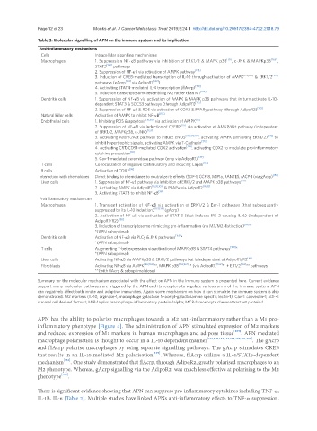
Table 3. Molecular signalling of APN on the immune system and its implication
Anti-inflammatory mechanisms
Cells Intracellular siganlling mechanisms
Macrophages 1. Suppression NF-κB pathway via inhibition of ERK1/2 & MAPK p38 [131] , c-JNK & MAPKp38 [168] ,
STAT3 [168] pathways
2. Suppression of NF-κB via activation of AMPK pathway [128]
3. Induction of CREB-mediated transcription of IL-10 through activation of AMPK [129,130] & ERK1/2 [129]
pathways (gAcrp [130] via AdipoR1 [130] )
4. Activating STAT4-mediated IL-4 transcription (flAcrp) [130]
5. Induction transcriptosome resembling M2 rather than M1 [169]
Dendritic cells 1. Suppression of NF-κB via activation of AMPK & MAPK p38 pathways that in turn activate IL-10-
dependent STAT3 & SOCS3 pathways (through AdipoR1) [145]
2. Suppression of NF-κB & ROS via activation of COX2 & PPARg pathway (through AdipoR2) [145]
Natural killer cells Activation of AMPK to inhibit NF-κB [155]
Endothelial cells 1. Inhibiting ROS & apoptosis [18,150] via activation of AMPK [18]
2. Suppression of NF-κB via induction of C/EBP [151] , via activation of AMKP/Akt pathway (independent
of ERK1/2, MAPKp38, c-JNK) [153]
3. Activating AMPK/Akt pathway to induce eNOS [140,170,171] , activating AMPK (inhibiting ERK1/2) [172] to
inhibit hypertrophic signals, activating AMPK via T-Cadherin [173]
4. Activating CRT/CD91-mediated COX2 activation [174] , activating COX2 to modulate pro-inflammatory
cytokine production [18]
5. Cav-1-mediated ceramidase pathway (only via AdipoR1) [147]
T cells Co-localization of negative costimulatory and inducing Capas [156]
B cells Activation of COX2 [36]
Interaction with chemokines Direct binding to chemokines to neutralize its effects (SDF-1, CCF18, MIP1a, RANTES, MCP-1 (via gAcrp) [157]
Liver cells 1. Suppression of NF-κB pathway via inhibition of ERK1/2 and MAPK p38 pathways [175]
2. Activating AMPK via AdipoR1 [19,20,103] & PPAPα via AdipoR2 [19,20]
3. Activating STAT3 to inhibit NF-κB [161]
Proinflammatory mechanisms
Macrophages 1. Transient activation of NF-κB via activation of ERK1/2 & Egr-1 pathways (that subsequently
suppressed by its IL-10 induction) [133,138] (gAcrp)
2. Activation of NF-κB via activation of STAT-3 (that induces IRS-2 causing IL-6) (independent of
AdipoR1/R2) [158]
3. Induction of transcriptosome mimicking pro-inflammation (no M1/M2 distinction) [160] *
*(APN suboptimal)
Dendritic cells Activation of NF-κB via PLCg & JNK pathways [159] *
*(APN suboptimal)
T cells Augmenting T-bet expression via activation of MAPKp38 & STAT4 pathways [160] *
*(APN suboptimal)
Liver cells Activating NF-κB via MAPKp38 & ERK1/2 pathways but is independent of AdipoR1/R2 [161]
Fibroblasts Activating NF-κB via AMPK [162,163] **, MAPK p38 [162,163] ** (via AdipoR1) [162] ** + ER1/2 [163] ** pathways
**(with flAcrp & suboptimal dose)
Summary for the molecular mechanism associated with the effect on APN in the immune system is presented here. Current evidence
support many molecular pathways are triggered by the APN and its receptors to regulate various arms of the immune system. APN
can negatively affect both innate and adaptive immunities. Again, some mechanism on how it can stimulate the immune system is also
demonstrated. M2 markers (IL-10, arginase-1, macrophage galactose N-acetyl-galactosamine specific lectin-1). Cav-1: caoveolin-1; SDF-1:
stromal cell derived factor-1; MIP-1alpha: macrophage-inflammatory protein-1alpha; MCP-1: monocyte chemoattractant protein-1
APN has the ability to polarise macrophages towards a M2 anti-inflammatory rather than a M1 pro-
inflammatory phenotype [Figure 4]. The administration of APN stimulated expression of M2 markers
and reduced expression of M1 markers in human macrophages and adipose tissue [169] . APN mediated
macrophage polarisation is thought to occur in a IL-10 dependent manner [127,129,131,135,138,169,181,182] . The gAcrp
and flAcrp polarise macrophages by using separate signalling pathways. The gAcrp stimulates CREB
that results in an IL-10 mediated M2 polarisation [129] . Whereas, flAcrp utilizes a IL-4/STAT6-dependent
mechanism [182] . One study demonstrated that flAcrp, through AdipoR2, greatly polarised macrophages to an
M2 phenotype. Whereas, gAcrp signalling via the AdipoR2, was much less effective at polarising to the M2
phenotype [182] .
There is significant evidence showing that APN can suppress pro-inflammatory cytokines including TNF-α,
IL-1B, IL-6 [Table 2]. Multiple studies have linked APNs anti-inflammatory effects to TNF-α suppression.