
Page 292 - Read Online
P. 292
Page 6 of 10 Bracht et al. J Cancer Metastasis Treat 2019;5:22 I http://dx.doi.org/10.20517/2394-4722.2018.111
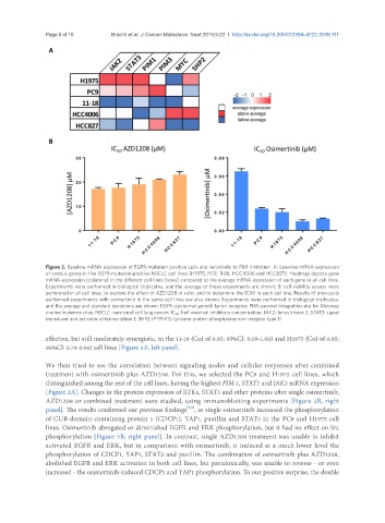
Figure 2. Baseline mRNA expression of EGFR-mutation positive cells and sensitivity to PIM inhibition. A: baseline mRNA expression
of various genes in five EGFR-mutation-positive NSCLC cell lines (H1975, PC9, 11-18, HCC4006 and HCC827). Heatmap depicts gene
mRNA expression (columns) in the different cell lines (rows) compared to the average mRNA expression of each gene in all cell lines.
Experiments were performed in biological triplicates, and the average of these experiments are shown; B: cell viability assays were
performed in all cell lines, to explore the effect of AZD1208 in vitro, and to determine the IC50 in each cell line. Results of previously
performed experiments with osimertinib in the same cell lines are also shown. Experiments were performed in biological triplicates,
and the average and standard deviations are shown. EGFR: epidermal growth factor receptor; PIM: proviral integration site for Moloney
murine leukemia virus; NSCLC: non-small cell lung cancer; IC 50 : half maximal inhibitory concentration; JAK2: Janus kinase 2; STAT3: signal
transducer and activator of transcription 3; SHP2 (PTPN11): tyrosine-protein phosphatase non-receptor type 11
effective, but still moderately synergistic, in the 11-18 (CoI of 0.85; 95%CI: 0.69-1.00) and H1975 (CoI of 0.85;
95%CI: 0.74-0.96) cell lines [Figure 3A, left panel].
We then tried to see the correlation between signaling nodes and cellular responses after combined
treatment with osimertinib plus AZD1208. For this, we selected the PC9 and H1975 cell lines, which
distinguished among the rest of the cell lines, having the highest PIM-1, STAT3 and JAK2 mRNA expression
[Figure 2A]. Changes in the protein expression of RTKs, STAT3 and other proteins after single osimertinib,
AZD1208 or combined treatment were studied, using immunoblotting experiments [Figure 3B, right
[6,8]
panel]. The results confirmed our previous findings , as single osimertinib increased the phosphorylation
of CUB-domain-containing protein 1 (CDCP1), YAP1, paxillin and STAT3 in the PC9 and H1975 cell
lines. Osimertinib abrogated or diminished EGFR and ERK phosphorylation, but it had no effect on Src
phosphorylation [Figure 3B, right panel]. In contrast, single AZD1208 treatment was unable to inhibit
activated EGFR and ERK, but in comparison with osimertinib, it induced at a much lower level the
phosphorylation of CDCP1, YAP1, STAT3 and paxillin. The combination of osimertinib plus AZD1208,
abolished EGFR and ERK activation in both cell lines, but paradoxically, was unable to reverse - or even
increased - the osimertinib-induced CDCP1 and YAP1 phosphorylation. To our positive surprise, the double