
Page 198 - Read Online
P. 198
Page 4 of 13 Suominen et al. J Cancer Metastasis Treat 2019;5:14 I http://dx.doi.org/10.20517/2394-4722.2018.64
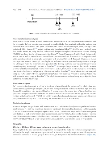
Figure 1. Dosing schedule of the doxorubicin (DOX) + zoledronic acid (ZOL) group
Histomorphometric analyses
After fixation in 10% neutral buffered formalin and decalcification in 10% ethylenediaminetetraacetic acid
for two weeks, the bone samples were processed to paraffin blocks. Four µm thick midsagittal sections were
obtained from the left hind limb (tibia and femur) and stained with hematoxylin, eosin, Orange G and
[6]
[16]
phloxine B (HE + Orange G) , tartrate-resistant acid phosphatase (TRAP) , Ki-67 (primary antibody clone
SP6, Nordic BioSite AB, Täby, Sweden) and terminal deoxynucleotidyl transferase dUTP nick end labeling
(TUNEL) methods (in situ cell death detection kit, AP™, Roche Diagnostics GmbH, Basel, Switzerland).
Tumor area as well as trabecular and cortical bone areas were analyzed from the HE + Orange G stained
slides as follows: first, micrographs were taken with a Leica DM4000 B Research Microscope (Leica
Microsystems, Wetzlar, Germany), then brightness and contrast were optimized using the same settings
for all images. Next, the tumor area was quantitated by drawing and bone area was quantitated by color
[8]
tresholding using MetaMorph™ software as described . Areas expanding 5 mm from the articular surfaces
of femur and tibia were analyzed. From a TRAP stained section, the number of osteoclasts at the tumor-bone
[18]
interface in distal femur and proximal tibia was counted as described , the whole section was analyzed
using the MetaMorph™ software. Apoptotic cells in tumor were manually counted as TUNEL stained cells
[6]
with apoptotic morphology as described . The whole tumor area was analyzed using a 20× objective Leica
DM4000 B Research Microscope.
Biomarker analyses
2+
Ca concentration (corrected to pH 7.4 by the internal algorithm of the instrument) in whole blood was
determined using a blood gas analyzer (ABL835 Flex blood gas analyzer, Radiometer Medical ApS, Bronshoj,
Denmark) immediately after terminal bleeding. A comparison to the normal level of ionized calcium was
performed using the values obtained from five intact animals of the same strain, sex and age. Serum tartrate-
resistant acid phosphatase 5b (TRACP 5b) activity as a marker of osteoclast number was measured in serum
samples obtained on days 1, 9, 17 and 24 using the MouseTRAP kit (IDS, Boldon, UK).
Statistical analyses
Statistical analysis was performed with SPSS (version 14.0). All statistical analyses were performed as two-
sided tests and P < 0.05 was considered statistically significant. The normality of residuals and homogeneity
of variances were examined using Kolmogorov-Smirnov and Levene’s tests, respectively. If these assumptions
were fulfilled as such or after appropriate transformation, one-way ANOVA followed by Dunnett t-test was
used. If the assumptions were not fulfilled even after transformation, the non-parametric Kruskal-Wallis test
followed by Mann-Whitney U-test was used. Fischer Exact test was used for comparison of proportions.
RESULTS
Effects of DOX and ZOL on body weight and tumor characteristics
Body weight of the mice decreased during the last five days of the study due to the disease progression.
Although the weight loss was more pronounced in the DOX treated groups, statistically significant
differences compared to vehicle group in the body weight change from day 0 were not observed [Figure 2B].