
Page 43 - Read Online
P. 43
Kaneko et al. Hyperostosis of metastatic adenocarcinoma by gefitinib
complaining of backache that had developed two DISCUSSION
weeks before. She had no history of smoking. Physical
examination revealed no significant findings. However, A phenomenon, so called osteoblastic flare, has
chest X-ray showed a mass shadow at the left upper originally been recognized as a transient increased
lung field, and computed tomography (CT) confirmed uptake of radiotracer of bone scintigraphy. [1,7] However,
a tumor of 4.5 cm in diameter with irregular margins ALP appeared to replace later because of infrequent
at the left upper lobe. The tumor was accompanied by use of bone scan, rapid and easy application of
[6]
ground glass-like consolidation [Figure 1A]. Osteolytic ALP, and coincident fluctuation of both. Our patient
lesions of the fourth thoracic spine [Figure 2A] and demonstrated a rapid improvement of bone pain and
the fourth left rib [Figure 2B] were also shown. tumor regression by gefitinib. This study might support
[3]
Laboratory data demonstrated elevation of serum the previous report by Arai et al. suggesting a
carcinoembryonic antigen (CEA) [Table 1]. ALP was favorable response to EGFR-TKI in case of ALP flare.
also elevated at 379 IU/L (normal range from 104 to It is of interest that Shimazaki et al. first observed
[8]
338). CT-guided needle biopsy was carried out and ALP flare-like phenomenon in a patient with multiple
the acquired specimen was pathologically diagnosed myeloma who received bortezomib for recurrent
as adenocarcinoma. The tumor was also found to carry massive bone lesions. Their patient showed a transient
a mutation of EGFR (L858R). Treatment with gefitinib ALP-3 increase without disease progression. Recent
began and her subjective symptom was relieved extreme efficacy of novel therapeutic agents might
quickly within several days.
Table 1: Laboratory data on admission
On treatment day 13, ALP increased to 952 IU/L, about Inspection item Value
2.5 times the pretreatment level. Serum transaminases White blood cells 10,790 μL 4
were also simultaneously elevated (AST 101 Red blood cells 456 × 10 μL
Hemoglobin
13.6 g/dL
and ALT 168), suggesting gefitinib-induced liver Hematocrit 40.5%
4
dysfunction. Serum Ca remained within normal limits. Platelets 38.5 × 10 μL
137.9 ng/mL (0-5.0)*
Carcinoembryonic entigen
Electrophoretic analysis showed that ALP-isozyme 1 Sialyl Lewis-X antigen 110 ng/mL (< 38.0)*
accounted for 8.6%, ALP-2 56.3%, and ALP-3 35.1%, Alkaline phosphatase 379 IU/L (104-338)*
respectively. Although bone-derived ALP-3 was seen Lactate dehydrogenase 468 IU/L (108-221)*
Asparate aminotransferase
25 IU/L
to increase, ALP-2 liver-derived isozyme had the larger Alanine aminotranferase 14 IU/L
increase, probably because of simultaneous drug- Total bilirubin 0.7 mg/dL
Leucine aminopeptidase
59 IU/L
induced liver injury. Gamma glutamyl transpeptidase 37 IU/L
Albumin 3.9 g/dL
Blood urea nitrogen 11.5 mg/dL
Since the significance of elevated ALP was unclear, Creatinine 0.76 mg/dL
another CT was carried out on treatment day 38. Uric acid 4.8 mg/dL
Results showed the primary pulmonary tumor was Calcium 10.2 mg/dL
142 mEq/L
Natrium
reduced to 3 cm in diameter [Figure 1B]. Previously Kalium 4.2 mEq/L
osteolytic lesions had become osteosclerotic. Lesion Chloride 103 mEq/L
C-reactive protein
0.10 mg/dL
sizes are evidenced excessive growth, larger than HBsAg negative
the size of the original bones [Figure 2C and D]. ALP HBsAb negative
negative
HBcAb
gradually decreased and liver dysfunction regressed HCVAb negative
[Figure 3]. CEA also decreased to 34.7 ng/mL, about *Abnormal data are in bold. Their normal ranges are indicated in
one fourth of the maximum value [Figure 3]. the following parentheses
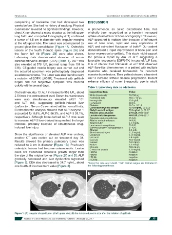
Figure 1: (A) Irregular shaped tumor at left upper lobe; (B) the tumor reduced in size after the initiation of gefitinib
Journal of Cancer Metastasis and Treatment ¦ Volume 3 ¦ February 23, 2017 35