
Page 39 - Read Online
P. 39
Warawdekar et al. CTCs from patients with metastatic breast cancer
nucleus. Figure 6 shows representative images of the presence of CTCs in samples.
two CTCs characterized as EpCAM and CK positive,
with a well-defined nucleus and were negative for the DISCUSSION
leucocyte antigen, CD45. Imaging further confirmed We propose a workable method for the isolation and
enumeration of CTCs wherein a two-tier protocol
of cell isolation with an initial separation based on
density gradient centrifugation, followed by EpCAM
immunomagnetic-positive double enrichment has been
described and adopted. The enriched fraction of tumour
cells is further divided, one analyzed for enumeration
of CTCs using flow cytometry based on large size of
tumor cells, with the presence of CK, EpCAM, and
the absence of CD45. Dissimilar expression levels of
EpCAM could compromise the detection of CTCs, [26,27]
hence, the initial standardization for flow and QRT-
PCR analysis was done with cell spiking of cancer
cells from different breast cancer subtypes to assess
possible differences. Tumor cells from the different
subtypes exhibited similar staining to EpCAM and CK
antibodies and could be clearly distinguished from
Jurkat cells. As described for serial dilutions [Figure 2]
recovery and linearity showed reproducibility and were
highly consistent across independent experiments. A
positive correlation was observed between recovered
tumor events and expected tumor events. Based on
the serial dilution assay, the percentage of tumor
cells recovered was not significantly different from the
percentage of tumor cells expected.
The flow cytometry protocol was validated with blood
samples obtained from patients with metastatic
breast cancer. Eighteen of these patients were with
tumor grade II-III and 17 were diagnosed with verified
metastasis. Clinical characteristics are as described
in Supplementary Table 1. Median age was 50 years
(range: 25-76 years). All, except two, were diagnosed
as invasive ductal carcinoma, the most common type
of breast cancer. Hormone receptor status showed
10 (45%) were ER-positive, 6 (27%), ER-negative,
equal number 9 (41%) PR-positive and PR-negative,
3 (14%) Her2-positive, and 13 (60%) Her2-negative.
CTCs showed a range of 1-85 per 10 mL of blood, with
an average of 23.35 ± 22.85. Twenty healthy women
volunteers were also included in this validation, with
values ranging from 0-14 per 10 mL of blood, with an
average of 5 ± 4. A cut-off of 10 and above has been
selected, based on these results.
For clinical samples where CTCs were higher,
captured cells were subjected to morphological
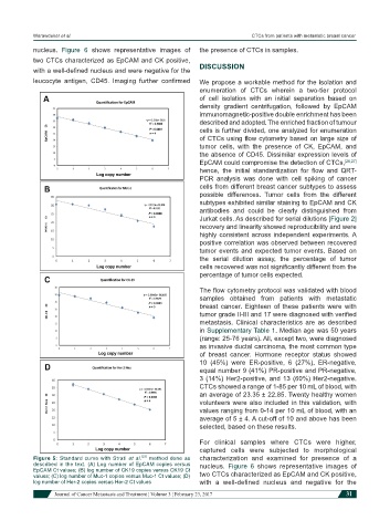
Figure 5: Standard curve with Strati et al. [23] method done as characterization and examined for presence of a
described in the text. (A) Log number of EpCAM copies versus nucleus. Figure 6 shows representative images of
EpCAM Ct values; (B) log number of CK19 copies versus CK19 Ct
values; (C) log number of Muc-1 copies versus Muc-1 Ct values; (D) two CTCs characterized as EpCAM and CK positive,
log number of Her-2 copies versus Her-2 Ct values with a well-defined nucleus and negative for the
Journal of Cancer Metastasis and Treatment ¦ Volume 3 ¦ February 23, 2017 31