
Page 85 - Read Online
P. 85
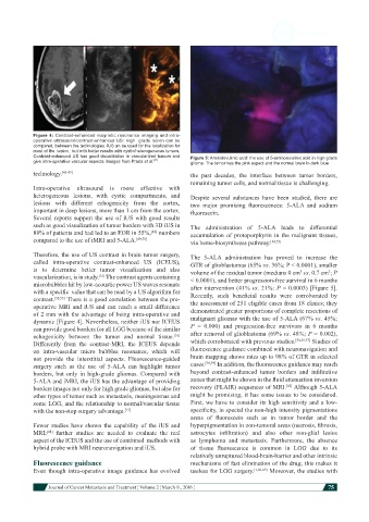
Figure 4: Contrast-enhanced magnetic resonance imaging and intra-
            operative ultrasound/contrast-enhanced US: High  grade lesion can be
            compared, between the technologies. iUS an be used for the localization for
            most of the  lesion,  but with better results with cystic/heterogeneous tumors.
            Contrast-enhanced US has good visualization in vascularized tumors and   Figure 5: Aminolevulinic acid: the use of 5-aminolevulinic acid in high grade
            give intra-operative vascular aspects. Images from Prada et al. [51]  glioma. The tumor has the pink aspect and the normal brain in dark blue
            technology. [45-47]                                the  past  decades,  the  interface  between  tumor  borders,
                                                               remaining tumor cells, and normal tissue is challenging.
            Intra-operative  ultrasound  is  more  effective  with
            heterogeneous lesions, with cystic compartments,  and   Despite  several  substances  have  been  studied,  there  are
            lesions with  different  echogenicity  from  the  cortex,   two  major  promising  fluorescences:  5-ALA  and  sodium
            important in deep lesions, more than 1 cm from the cortex.   fluorescein.
            Several reports support the use of iUS with good results
            such as good visualization of tumor borders with 3D iUS in   The  administration  of  5-ALA leads  to  differential
            88% of patients and had led to an EOR in 55%,  numbers   accumulation  of protoporphyrin in the malignant  tissues,
                                                  [48]
            compared to the use of iMRI and 5-ALA. [49,50]     via heme-biosyntheses pathway. [54,55]
            Therefore, the use of US contrast in brain tumor surgery,   The  5-ALA  administration  has  proved  to  increase  the
            called  intra-operative  contrast-enhanced  US  (ICEUS),   GTR of glioblastomas (65% vs. 36%; P < 0.0001), smaller
            is  to  determine  better  tumor  visualization  and  also   volume of the residual tumor (medians 0 cm  vs. 0.7 cm ; P
                                                                                                           3
                                                                                                  3
            vascularization, is in study.  The contrast agents containing   < 0.0001), and better progression-free survival in 6 months
                                 [51]
            microbubbles hit by low-acoustic power US waves resonate    after intervention (41% vs. 21%: P = 0.0003) [Figure 5].
            with a specific  value that can be read by a US algorithm for   Recently,  such  beneficial  results  were  corroborated  by
            contrast. [52,53]  There is a good correlation between the pre-  the assessment of 251 eligible cases from 18 clinics; they
            operative MRI and iUS and can reach a small difference   demonstrated greater proportions of complete resections of
            of 2 mm with the advantage of being intra-operative and
            dynamic [Figure 4]. Nevertheless, neither iUS nor ICEUS   malignant gliomas with the use of 5-ALA (67% vs. 45%;
            can provide good borders for all LGG because of the similar   P  =  0.000)  and  progression-free  survivors  in  6  months
            echogenicity  between  the  tumor  and  normal  tissue.    after removal of glioblastoma (69% vs. 48%; P = 0.002),
                                                         [51]
                                                                                                [54,56,57]
            Differently  from  the  contrast-MRI,  the  ICEUS  depends   which corroborated with previous studies.   Studies of
            on  intra-vascular  micro  bubbles  resonance,  which  will   fluorescence guidance combined with neuronavigation and
            not  provide  the  interstitial  aspects.  Fluorescence-guided   brain mapping shows rates up to 98% of GTR in selected
            surgery such as the use of 5-ALA can highlight tumor   cases. [58,59]  In addition, the fluorescence guidance may reach
            borders, but  only in  high-grade gliomas.  Compared  with   beyond  contrast-enhanced  tumor  borders  and  infiltrative
            5-ALA and iMRI, the iUS has the advantage of providing   zones that might be shown in the fluid attenuation inversion
                                                                                             [42]
            borders images not only for high grade gliomas, but also for   recovery (FLAIR) sequences of MRI.  Although 5-ALA
            other types of tumor such as metastasis, meningeomas and   might be promising, it has some issues to be considered.
            some LGG, and the relationship to normal/vascular tissue   First, we have to consider its high sensitivity and a low-
            with the non-stop surgery advantage. [51]          specificity, in special the non-high intensity pigmentations
                                                               areas  of  fluorescein  such  as  in  tumor  border  and  the
            Fewer  studies  have  shown  the  capability  of  the  iUS  and   hyperpigmentation in non-tumoral areas (necrosis, fibrosis,
            MRI;   further  studies  are  needed  to  evaluate  the  real   astrocytes  infiltration)  and  also  other  non-glial  lesios
                [48]
            aspect of the ICEUS and the use of combined  methods with   as lymphoma  and metastasis.  Furthermore,  the absence
            hybrid probe with MRI neuronavigation and iUS.     of  tissue  fluorescence  is  common  in  LGG  due  to  its
                                                               relatively unruptured blood-brain-barrier and other intrinsic
            Fluorescence guidance                              mechanisms of fast elimination of the drug; this makes it
            Even  though  intra-operative image  guidance has  evolved   useless for LGG surgery. [1,60-63]  Moreover, the studies with


                        Journal of Cancer Metastasis and Treatment ¦ Volume 2 ¦ March 11, 2016 ¦           75