
Page 483 - Read Online
P. 483
Gajjar et al. ERCC1 in colorectal cancer
are outlined in Table 1. The median age was 55 years.
There was a considerable proportion (54%) of elderly
and dominance of male patients (60%). The incidence
of patients having colon cancer was higher (56%) as
compared to rectal cancer (44%). Higher occurrence
of early stage patients (60%) was observed. Complete
follow-up details were obtained in 80% (40/50) and
were included for OS analysis. Amongst these 40, 4
died due to disease and hence were not included for
the RFS analysis. Therefore, 36/40 CRC patients were
considered for RFS analysis from which 2 patients
developed recurrence [Table 1].
Incidence of ERCC1 codon 118 C/T
polymorphism
Three types of genotypes have been identified for
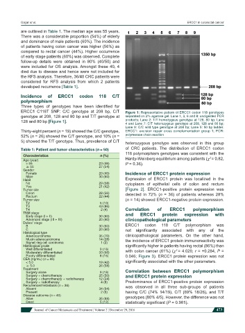
ERCC1 C118T SNP: C/C genotype at 208 bp, C/T Figure 1: Representative pattern of ERCC1 codon 118 genotypes
genotype at 208, 128 and 80 bp and T/T genotype at separated on 2% agarose gel. Lane 1, 3, 6 and 8: undigested PCR
128 and 80 bp [Figure 1]. products; Lane 2: T/T homozygous genotype at 128, 80 bp; Lane
4 and Lane 7: C/T heterozygous genotype at 208, 128 and 80 bp;
Lane 9: C/C wild type genotype at 208 bp; Lane 5: 50 bp ladder.
Thirty-eight percent (n = 19) showed the C/C genotype, ERCC1: excision repair cross complementation group 1; PCR:
52% (n = 26) showed the C/T genotype, and 10% (n = polymerase chain reaction
5) showed the T/T genotype. Thus, prevalence of C/T heterozygous genotype was observed in this group
Table 1: Patient and tumor characteristics (n = 50) of CRC patients. The distribution of ERCC1 codon
Characteristics n (%) 118 polymorphism genotypes was consistent with the
2
Age (year) Hardy-Weinberg equilibrium among patients (χ = 0.82,
< 55 23 (46) P = 0.36).
≥ 55 27 (54)
Gender
Female 20 (40) Incidence of ERCC1 protein expression
Male 30 (60) Expression of ERCC1 protein was localized in the
Habit
No 29 (58) cytoplasm of epithelial cells of colon and rectum
Yes 21 (42)
Tumor site [Figure 2]. ERCC1-positive protein expression was
Colon 28 (56) detected in 72% (n = 36) of patients whereas 28%
Rectum 22 (44)
Tumor size (n = 14) showed ERCC1-negative protein expression.
T2 5 (10)
T3 43 (86)
T4 2 (4) Correlation of ERCC1 polymorphism
TNM stage and ERCC1 protein expression with
Early stage (I + II) 30 (60)
Advanced stage (III + IV) 20 (40) clinicopathological parameters
Dukes’ stage ERCC1 codon 118 C/T polymorphism was
B 30 (60)
C 20 (40) not significantly associated with any of the
Histological type
Adenocarcinoma 35 (70) clinicopathological parameters. On the other hand,
Mucin adenocarcinoma 14 (28) the incidence of ERCC1 protein immunoreactivity was
Signet ring cell carcinoma 1 (2)
Histological grade significantly higher in patients having rectal (86%) than
Well differentiated 9 (18) with colon cancer (61%) (χ = 4.020, r = +0.284, P =
2
Moderately differentiated 33 (66)
Poorly differentiated 8 (16) 0.046; Figure 3). ERCC1 protein expression was not
CEA (ng/mL) (n = 45)
< 5.0 19 (42) significantly associated with the other parameters.
≥ 5.0 26 (58)
Treatment Correlation between ERCC1 polymorphism
Surgery alone 9 (18)
Surgery + chemotherapy 25 (50) and ERCC1 protein expression
Surgery + chemotherapy + radiotherapy 12 (24)
Surgery + radiotherapy 4 (8) Predominance of ERCC1-positive protein expression
Recurrence/metastasis (n = 36) was observed in all three sub-groups of patients
Absent 34 (94)
Present 2 (6) having C/C (74% 14/19), C/T (69% 18/26), and T/T
Disease outcome (n = 40) genotypes (80% 4/5). However, the difference was not
Alive 35 (88)
Dead 5 (12) statistically significant (P = 0.981).
Journal of Cancer Metastasis and Treatment ¦ Volume 2 ¦ December 29, 2016 473