
Page 69 - Read Online
P. 69
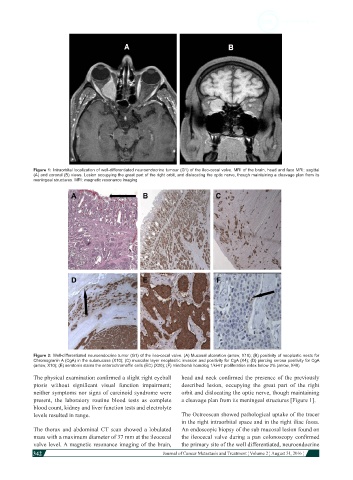
Figure 1: Intraorbital localization of well-differentiated neuroendocrine tumour (G1) of the ileo-cecal valve. MRI of the brain, head and face MRI: sagittal
            (A) and coronal (B) views. Lesion occupying the great part of the right orbit, and dislocating the optic nerve, though maintaining a cleavage plan from its
            meningeal structures. MRI: magnetic resonance imaging


            Figure 2: Well-differentiated neuroendocrine tumor (G1) of the ileo-cecal valve. (A) Mucosal ulceration (arrow, X10); (B) positivity of neoplastic nests for
            Chromogranin A (CgA) in the submucosa (X10); (C) muscolar layer neoplasitic invasion and positivity for CgA (X4); (D) piercing serosa positivity for CgA
            (arrow, X10); (E) serotonin stains the enterochromaffin cells (EC) (X20); (F) mindbomb homolog 1/ki-67 proliferation index below 2% (arrow, X40)
            The physical examination confirmed a slight right eyeball   head and neck confirmed the presence of the previously
            ptosis  without  significant  visual  function  impairment;   described lesion, occupying the great part of the right
            neither symptoms nor signs of carcinoid syndrome were   orbit and dislocating the optic nerve, though maintaining
            present, the laboratory  routine blood tests as complete   a cleavage plan from its meningeal structures [Figure 1].
            blood count, kidney and liver function tests and electrolyte
            levels resulted in range.                         The Octreoscan showed pathological uptake of the tracer
                                                              in the right intraorbital space and in the right iliac fossa.
            The  thorax  and  abdominal  CT scan  showed a  lobulated   An endoscopic biopsy of the sub mucosal lesion found on
            mass with a maximum diameter of 37 mm at the ileocecal   the ileocecal valve during a pan colonoscopy confirmed
            valve level. A magnetic resonance imaging of the brain,   the primary site of the well differentiated, neuroendocrine
            342
                                                                                                                   Journal of Cancer Metastasis and Treatment ¦ Volume 2 ¦ August 31, 2016 ¦