
Page 66 - Read Online
P. 66
episodes occurred and hypoglycemia was biochemically
confirmed with an average 7 h serum glucose concentration
of 45 mg/dL (normal range 80-120 mg/dL). Hypoglycemic
episodes fulfilled the Whipple’s triad, characterized by
signs and symptoms of hypoglycemia, evidence of low
plasma glucose (< 55 mg/dL) concentration and resolution
of signs and symptoms after glucose administration. Liver,
renal and thyroid profiles were within the normal limits.
An insulinoma was suspected and a 72 h fasting test was
performed with assessment of glycemia at the beginning and
every 4 h. Serum insulin and C-Peptide concentrations were
also assessed at the beginning and in case of biochemical
and/or clinical hypoglycemia. Serum concentrations of
glucose, insulin and C-peptide were measured by standard
methods by using commercially available kits. During the
test hypoglycemia occurred after 9 h (glucose 40 mg/dL).
However, the insulin/glucose ratio was 0.1, revealing an
appropriate insulin secretion. Moreover, a focal lesion
within the pancreas was detected by endoscopic ultrasound
(EUS), therefore an insulinoma was suspected. However,
the evaluation of pituitary function with growth hormone-
releasing hormone (GHRH) plus arginine test pointed
out a growth hormone (GH) deficiency and magnetic
resonance imaging (MRI) of the pituitary region revealed
a partial empty sella. No other pituitary abnormalities
were observed. An abdominal contrast-enhanced CT
confirmed a nodular area of 18 mm × 12 mm in the body
of pancreas, with altered contrast enhancement. An 111In-
DTPA-D-Phe1 octreotide scintigraphy (Octreoscan)
highlighted a focal epigastric uptake, corresponding to the
pancreatic nodule. Surprisingly a EUS-guided fine-needle
biopsy of the pancreatic lesion resulted in a cytological
diagnosis of moderately differentiated adenocarcinoma.
Therefore, the patient underwent surgery. Histology
and immunohistochemistry of the specimen revealed
a well-differentiated pNET, with Ki67 index of 1%.
Immunostaining for chromogranin-A and synaptophysin
was positive, while insulin immunostaining was negative.
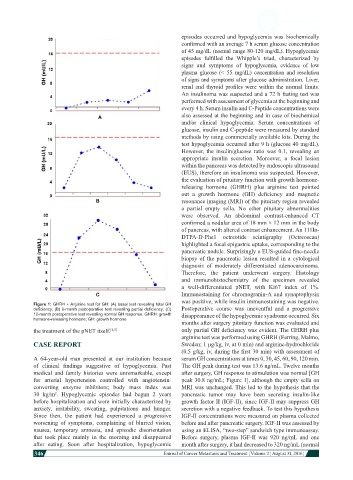
Figure 1: GHRH + Arginine test for GH: (A) basal test revealing total GH
deficiency; (B) 6-month postoperative test revealing partial deficiency; (C) Postoperative course was uneventful and a progressive
12-month postoperative test revealing normal GH response. GHRH: growth disappearance of the hypoglycemic syndrome occurred. Six
hormone-releasing hormone; GH: growth hormone
months after surgery pituitary function was evaluated and
the treatment of the pNET itself. [4,5] only partial GH deficiency was evident. The GHRH plus
arginine test was performed using GHRH (Ferring, Malmo,
CASE REPORT Sweden; 1 μg/kg, iv, at 0 min) and arginine-hydrochloride
(0.5 g/kg, iv, during the first 30 min) with assessment of
A 64-year-old man presented at our institution because serum GH concentrations at times 0, 30, 45, 60, 90, 120 min.
of clinical findings suggestive of hypoglycemia. Past The GH peak during test was 13.6 ng/mL. Twelve months
medical and family histories were unremarkable, except after surgery, GH response to stimulation was normal [GH
for arterial hypertension controlled with angiotensin- peak 30.8 ng/mL; Figure 1], although the empty sella on
converting enzyme inhibitors; body mass index was MRI was unchanged. This led to the hypothesis that the
30 kg/m . Hypoglycemic episodes had begun 2 years pancreatic tumor may have been secreting insulin-like
2
before hospitalization and were initially characterized by growth factor II (IGF-II), since IGF-II may suppress GH
anxiety, irritability, sweating, palpitations and hunger. secretion with a negative feedback. To test this hypothesis
Since then, the patient had experienced a progressive IGF-II concentrations were measured on plasma collected
worsening of symptoms, complaining of blurred vision, before and after pancreatic surgery. IGF-II was assessed by
nausea, temporary amnesia, and episodic disorientation using an ELISA, “two-step” sandwich type immunoassay.
that took place mainly in the morning and disappeared Before surgery, plasma IGF-II was 920 ng/mL and one
after eating. Soon after hospitalization, hypoglycemic month after surgery, it had decreased to 320 ng/mL (normal
346
Journal of Cancer Metastasis and Treatment ¦ Volume 2 ¦ August 31, 2016 ¦