
Page 81 - Read Online
P. 81
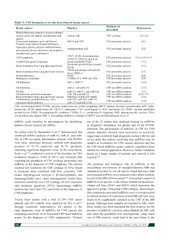
Table 3: CSF biomarkers for the detection of brain cancer
                                                                            Method of
            Brain cancer                        Marker                      detection            References
            Medulloblastoma, primitive neuroectodermal
            tumors, germ cell tumors, ependymoma and   Cancer cells         CSF cytology           [12-14]
            glioma
            Intracranial malignant germ cell tumors  bHCG and AFP           CSF proteomic analysis  [41]
            Pediatric brain tumors (medulloblastoma ,
            high-grade glioma, atypical rhabdoid tumor,   Apolipoprotein A-II  CSF proteomic analysis  [72]
            astrocytoma, plexus carcinoma and anaplastic
            ependymoma, germ cell tumor)
            CNS lymphoma                        CD27, AT III, chemoattractant,   CSF proteomic analysis  [55,57,107-111]
                                                CXCL13, CXCL12 and IL10
            Cerebral low-grade lymphoma         Immunoglobulin G IgG        CSF proteomic analysis  [112]
            Brain metastases from lung adenocarcinoma  Epidermal growth factor receptor   CSF proteomic analysis  [113]
                                                EGFR
            Brain metastases from lung and breast cancers  VEGF and stromal cell derived   CSF proteomic analysis  [73]
                                                factor (SDF)-1
            Medulloblastoma                     PGD2                        CSF proteomic analysis  [60]
            Meningeal carcinomas                CYFRA 21-1, NSE and CEA     CSF proteomic analysis  [70]
            Glioblastoma                        MIC-1 GDF15                 CSF proteomic analysis  [114]

            Glioblastoma                        miR-21 and miR-15b          CSF microRNA analysis   [115]
            PCNSL                               miR-19, miR-21, and miR-92a  CSF microRNA analysis  [115]
            Glioblastoma and brain metastasis   miR-10b and miR-21          CSF microRNA analysis  [116-117]
            Brain metastases from lung and breast cancers  Members of miR-200 family  CSF microRNA analysis  [116]
            Glioblastoma, medulloblastoma, brain   miR-935, miR-451, miR-711,   CSF microRNA analysis  [118]
            metastasis and lymphoma             miR-223 and miR-125b
           CSF: cerebrospinal fluid; PCNSL: primary central nervous system lymphoma; bHCG: human chorionic gonadotropin; AFP: alpha-
           fetoprotein; AT III: antithrombin III; CXCL13: chemokine C-X-C motif ligand 13; IL10: interleukin 10; VEGF: vascular endothelial
           growth factor; PGD2: Prostaglandin-D2 synthase; CYFRA 21-1: cytokeratin-19 fragment; NSE: neuron-specific enolase; CEA:
           carcinoembryonic antigen; MIC-1: macrophage inhibitory cytokine-1; GDF15: growth differentiation factor 15
           miRNA  could  therefore  be  advantageous  for  identifying   out of the 23 studies they analyzed  focused on miRNAs
           putative disease markers for DIPGs.                as diagnostic biomarkers for glioma and 10 for PCNSL
                                                              detection. The performance of miRNAs in CSF for CNS
           An earlier work by Baraniskin et al.  demonstrated that   cancers detection showed more correctness in sensitivity
                                         [77]
           combined miRNA analysis of miR-19, miR-21, and miR-  suggesting a relatively high diagnostic accuracy. By the end
           92a in CSF accurately discriminate patients with PCNSL   of the study the authors concluded that miRNAs may be
           from other neurologic disorders  controls with diagnostic   suitable as biomarkers for CNS cancers detection and that
           accuracy  of  95.7%  sensitivity  and  96.7%  specificity   the CSF based miRNAs assays could be considered more
           indicating significant diagnostic value. In the same theme,   reliable for clinical application. However, further validation
           Scott et al.  conducted a review of the literature on CNS   based  on a  larger  sample  of patients  and  controls  is still
                    [79]
           lymphoma  diagnosis  (1966  to  2011)  and  extracted  data   required. [81]
           regarding the usefulness of CSF cytology, proteomics and
           miRNAs in the diagnosis of CNS lymphoma. The authors   The  presence  and biological  role  of miRNAs in the
           reported low sensitivity for CSF cytology (2-32%) which   extracellular  environment  of  meddulloblastoma  MB  was
           is  increased  when  combined  with  flow  cytometry.  CSF   examined recently by our lab and we found that more than
           lactate  dehydrogenase  isozyme  5,  β2-microglobulin,  and   one thousand miRNAs were released in the culture-medium
           immunoglobulin  heavy chain  rearrangement  studies have   in each of the MB cell lines tested.  Among them a panel of
                                                                                         [82]
           improved sensitivity over CSF cytology (58-85%) but have   miRNAs were specific to the culture-medium of metastasis-
           only  moderate  specificity  (85%).  Interestingly  miRNA   related  cell  lines (D341 and D283) which represents the
           analysis has more than 95% specificity in the diagnosis of   aggressive group 3 and group 4 MB subtypes. Interestingly,
           CNS lymphoma.                                      three metastasis-associated miRNAs were over-represented
                                                              in culture-medium of metastasis-related MB cell lines were
           Twenty three studies with a total of 299 CNS cancer   found to be significantly enriched in the CSF of the MB
           patients  and 418 controls were analyzed  by Wei  et al.    patient. Although more samples are required to fully verify
                                                         [81]
           through systematic  meta-analysis  for articles  in the   these results, our work presented the first evidence for the
           topic  diagnostic  value  of  miRNAs for  CNS cancers  and   presence of miRNAs excreted extracellularly by MB cells
           comparing sensitivity of on blood-and CSF based miRNAs   and raises the possibility that investigations, using larger
           assays for the  diagnosis of CNS malignancies.  Thirteen   sets of MB samples, could lead in the near future to the

           182
                                                                                                                         Journal of Cancer Metastasis and Treatment ¦ Volume 2 ¦ May 18, 2016 ¦