
Page 77 - Read Online
P. 77
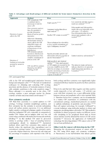
Table 2: Advantages and disadvantages of different methods for brain tumors biomarkers detection in the
           CSF
            Approach            Method                 Pros                       Cons
                                CSF cytoanalysis: CSF
                                is examined under a    Highly specific [12-14,16]  Low sensitivity and false negative
                                microscope to look for                            results are common [3,13,20,23,24]
                                cancer cells
                                Flow cytometry analysis:                          False negative and false positive
                                Have the potential to   Automated method that allows   results can occur (especially at low
                                                                 [14,25]
                                provide information    rapid analysis             cell counts, < 25 cells/uL).
            Detection of cancer   about cell surface protein   Smaller CSF volume is needed [9,23]  Poor differential ability between
            cells in the CSF    expression                                        mitoses and neoplastic cells is
                                                                                  reported
                                                                                        [14,23,25]
                                Other tools: Measuring
                                chromosomal content of
                                cancer cells in the CSF   These techniques have the ability
                                using DNA single cell   to detect genetic aberrations as a   Low sensitivity [26]
                                cytometry techniques   sign of malignancy location [26]
                                or fluorescence in-situ
                                hybridization
                                CSF proteomic analysis:
                                Systematic identification   Specific proteomic patterns can
                                and quantification of the   differentiate subtypes or grades of   Limited sensitivity and specificity [31]
                                complete complement of   specific brain tumors [27-30]
                                proteins in the CSF
            Detection of                               High specificity and chemical
            biochemical molecules                      stability [60,101]  Only small amounts
            secreted by cancers to                     of CSF samples are required for the  The unknown origin and factors
            the CSF             CSF microRNAs analysis:   detection of miRNAs in the CSF   influence their level of expression
                                Measuring microRNA
                                Profiling of CSF       offers the advantage of convenient   might impact their specificity as
                                                                                          [76,102-106]
                                                       repetitive monitoring of molecular   biomarkers
                                                       events happening in cancer in the
                                                       response to treatment [76]
           CSF: cerebrospinal fluid
           cells  in  the  CSF  and  morphological  similarities  between   both cytology and flow cytometry were significantly higher
           benign and malignant cells. [13,23,24]  The lack of standardized   compared with those found to be positive by flow cytometry
           techniques  for  obtaining  and  evaluating  CSF  cytology   alone. [23]
           specimens and the absence of molecular analysis of tumor
           cells certainly contributes to the wide sensitivity range.    It has to be said that both false negative and false positive
                                                         [3]
           Hence  although  it  is  currently  used  in  the  clinic,  CSF   results  (especially  at  low cell  counts,  < 25 cells/uL)  can
           cytology  remains  a poor surrogate  marker  for disease   occur with flow cytometry too, a poor differential ability
           response in brain cancer/metastasis involvement. [9,13]  between mitoses and neoplastic cells is also reported Table
                                                              2. Therefore before flow cytometry can be recommended
           Flow cytometry analysis                            in  a  routine  CSF  examination  in  combination  with  the
           CSF  fluid  flow  cytometry  is  a  useful  addition  to  CSF   conventional cytology, standardized protocols are needed
           cytology.  Cytology  examines  morphologic  patterns,  and   to  uniform  definitions  of  positivity  and  procedure. [14,23,25]
           flow  cytometry  has  the  potential  to  provide  information   Rare cell capture technology, for example, CellSearch  is
                                                                                                          ®
           about  cell  surface  protein  expression.  It  is  an  additional   a recent technique using molecular markers to detect and
           highly sensitive cytological technique capable of accurately   enumerate circulating tumor cells in the CSF. This method is
           detecting malignant CSF cells, especially in comparatively   established to detect prognostic marker on different cancer
           smaller  CSF  volume  and  in  samples  with  very  low  cell   cells circulating in the peripheral  blood such as breast
           counts when combined with multicolor fluorescent antibody   cancer and has recently attracted the interest of CSF cancer
           labelling. [9,23]   In  this  method  CSF  must  be  processed   researcher. [3,9]   However,  the  application  of  CellSearch
                                                                                                            ®
           similar  to cytology within 1 hour of sampling  however   technology for detecting primary CNS cancer cells in CSF
           centrifugation  should  be  minimized. [14,23]   Automated   has not been published yet.
           methods  allow  rapid  flow  cytometry  data  analysis  and
           thereby  reduce  the  significant  time  expenditures  used  in   Other tools for cancer cell detection in the CSF
           conventional cytology routine. [14,25]  Flow cytometry seems   Measuring the chromosomal  content of cancer  cells in
           to provide a higher sensitivity. However the cell count and   the  CSF,  using  DNA  single  cell  cytometry  techniques
           the percentage of neoplastic cells reported in the CSF by   or  fluorescence  in-situ hybridization  that  detects  genetic

           178
                                                                                                                         Journal of Cancer Metastasis and Treatment ¦ Volume 2 ¦ May 18, 2016 ¦