
Page 76 - Read Online
P. 76
It is accessible through lumbar puncture, a little invasive performing diagnostic postoperative CSF cytologic
procedure. Any cancer cells released by brain cancer bulk or evaluation. [9,16,17] Accurate cytopreparatory techniques
molecules that are actively secreted or passively diffused by are essential criteria for successful CSF microscopic
cancer cells are likely to disperse into the CSF and therefore evaluations. 7.5 mL of CSF are usually withdrawn and
can be detected. Hence CSF analysis is considered to be an immediately processed, as the cell counts can diminish by
important tool in the evaluation of CNS malignancies. This up to 50% within 2 h of collection. [4,18] CSF samples are
review discusses potential and limitations of CSF analyses then processed by centrifugation (CytospinÒ) at 800 g for
in brain cancer patients. 3-5 min, air-dried for 10-15 min and stained with May-
Grunwald Giemsa (MGG) stain solution for 10-15 min.
[19]
DETECTION OF CANCER CELLS IN THE Thin-layer preparation (ThinPrep) is a relatively new
CSF liquid-based cytology method which has been suggested
to better detect malignant cells in CSF from solid tumors
Primary CNS cancers and metastases are often located in by performing good preservation of cell morphologic
close proximity to ventricular surfaces or CSF cisterns. [9-11] features. During the ThinPrep analysis, the CSF cells are
Malignant cells derived from brain cancers reach the collected through high-precision filtration driven by fluid
leptomeninges by CSF spread or by direct extension mechanics and gently absorbed onto a glass slide by using
from the primary tumor itself e.g. medulloblastoma, electrochemical forces. The collected samples need to be
primitive neuroectodermal tumors, germ cell tumors, added to 10 mL preservation solution, mixed and stood
ependymoma, and glioma may be disseminated throughout for 15 min. Slides are fixed in 95% ethanol for 15 min and
the neuroaxis by the flow of the CSF. [12-14] Table 1 shows stained by standard Papanicolaou method. [19]
the particular incidence of malignant leptomeningeal
involvement in selected primary brain cancers. Currently CSF cytology, although indispensable, has many
microscopic evaluation of CSF is routinely performed in limitations Table 2. To start with it involves the pathological
CNS malignancies with frequent leptomeningeal spread, identification of abnormal cells in the CSF by Giemsa
such as medulloblastomas, PNET, pineoblastomas, germ- stain and clinicians must make judgments on the presence
cell tumors and CNS lymphoma. Cancer therapy and or absence of malignant cells. Hence, CSF cytological
[15]
prognosis of these groups of brain cancer are crucially analysis is a pure qualitative test that bears no quantification
determined by positive CSF cytology. [13,14] and lacks validation. [3,20] Another weakness is that because
the shedding of malignant cells into the CSF may occur
CSF cytoanalysis intermittently and in low numbers, inconsistent presence of
CSF cytology, in which CSF is prepared and examined cancer cells in the CSF should be expected. CSF specimens
under a microscope to look for cells, is currently considered may, therefore, fail to capture malignant cells representing
the gold standard for diagnosis of brain cancer with one of the major weaknesses of CSF cytology. It is therefore
leptomeningeal spread and metastatic cancer to the brain. recommended that CSF analysis should be repeated if
[14]
To achieve CSF cytology a sample can be obtained at the time initially negative. One of the drawbacks is while CSF
[21]
of tumor surgery or by lumbar or intracerebroventricular cytology is highly specific in detection of cancer cells, it
(ICV) reservoir puncture. However, lumbar CSF remains suffers from a lack of sensitivity. A retrospective meta-
[3]
the specimen of choice to detect malignant cells of primary analysis reported that CSF cytology sensitivity could be
[22]
CNS tumors. [12,16] To avoid false positive results due to as low as 45% depending on how many times the lumbar
sloughing of tumor cells at the time of surgery, a recovering puncture was repeated. False negative cytopathology is
interval of one to two weeks is currently suggested before common (10-20% of patients) because of the paucity of
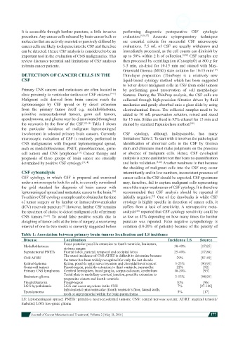
Table 1: Association between primary brain tumors localisation and LS incidence
Disease Localisation Incidence LS Source
Fossa posterior possible extension to fourth ventricle, brainstem,
Medulloblastoma cisterna magna 30-40% [17,85]
Supratentorial PNETs Frontal lobes, parietal, temporal and occipital lobes 25-40% [17,86]
CNS AT/RT The exact incidence of CNS AT/RT is difficult to determine because 29% [87-89]
the tumor has been widely recognized for only the last decade
Retinoblastoma Retina, possible optic nerve invasion and choroidal involvement 3-23% [90,91]
Germ-cell tumors Pineal-region, possible extension to third ventricle, suprasellar 22% [92]
Primary CNS lymphoma Cerebral hemisphers, basal ganglia, corpus callosum, cerebellum 10-20% [93]
Brainstem glioma Tectal plate to medullary cervical junction, possible extension to 3-13% [94,95]
prepontine cistern and fourth ventricle
Pinealoblastoma Pineal-region 10% [96]
LGG hypothalamic LGG can occur anywhere in the CNS 7% [97-100]
Ependymoma Infratentorial intraventricular (fourth ventricle’s floor, lateral walls, 5% [17]
roof) or supratentorial within the brain parenchyma
LS: leptomeningeal spread; PNETs: primitive neuroectodermal tumors; CNS: central nervous system; AT/RT: atypical teratoid/
rhabdoid; LGG: low-grade glioma
Journal of Cancer Metastasis and Treatment ¦ Volume 2 ¦ May 18, 2016 ¦ 177