
Page 50 - Read Online
P. 50
Page 8 of 14 Pinnamaneni et al. J Cancer Metastasis Treat 2021;7:7 I http://dx.doi.org/10.20517/2394-4722.2020.94
A C
B D
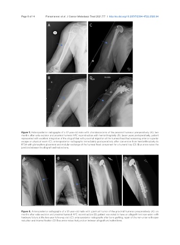
Figure 7. Anteroposterior radiographs of a 57-year-old male with: chondrosarcoma of the proximal humerus preoperatively (A); two
months after wide excision and proximal humeral APC reconstruction with hemiarthroplasty (B); Seven years postoperatively, patient
represented with excellent integration of the allograft but with proximal migration of the humeral head had worsening anterior superior
escape on physical exam (C); anteroposterior radiographs immediately postoperatively after conversion from hemiarthroplasty to
RTSA with glenosphere placement and modular exchange of the humeral head component for a humeral tray (D). Blue arrow notes the
junction between the allograft and native bone.
A B C D
Figure 8. Anteroposterior radiographs of a 55-year-old male with: giant cell tumor of the proximal humerus preoperatively (A); six
months after wide excision and proximal humeral APC reconstruction (B); patient was noted to have an allograft-host non-union with
hardware failure at the two-year follow-up visit (C); anteroposterior radiographs after bone grafting, repair of the non-union with open
reduction and internal fixation (D) Blue arrow notes that junction between allograft and native bone.