
Page 49 - Read Online
P. 49
Pinnamaneni et al. J Cancer Metastasis Treat 2021;7:7 I http://dx.doi.org/10.20517/2394-4722.2020.94 Page 7 of 14
A B
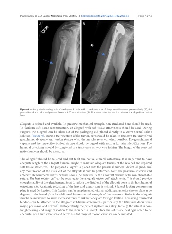
Figure 6. Anteroposterior radiographs of a 65-year-old male with: chondrosarcoma of the proximal humerus preoperatively (A); 4.5
years after wide excision and proximal humeral APC reconstruction (B). Blue arrow notes the junction between the allograft and native
bone.
allograft is ordered and available. To preserve mechanical strength, non-irradiated bone should be used.
To facilitate soft-tissue reconstruction, an allograft with soft-tissue attachments should be used. During
surgery, the allograft can be taken out of the packaging and placed directly in a warm normal saline
solution [Figure 9]. During the resection of the tumor, care should be taken to preserve the uninvolved
glenohumeral capsule and tendon stumps of all the muscles resected, when possible. The glenohumeral
capsule and the respective tendon stumps should be tagged with sutures for later identification. The
humeral osteotomy should be completed in a transverse or step-wise fashion. The length of the resected
native humerus should be measured.
The allograft should be tailored and cut to fit the native humeral osteotomy. It is important to have
adequate length of the allograft humeral height to maintain adequate tension of the retained and repaired
soft-tissue structures. The prepared allograft is placed into the proximal humeral defect, aligned, and
any modification of the distal cut of the allograft should be performed. Next, the posterior, inferior, and
anterior glenohumeral native capsule should be repaired to the allograft capsule with non-absorbable
suture. The host rotator cuff can be repaired to the allograft rotator cuff attachments. This should provide
enough stability of the glenohumeral joint to reduce the distal end of the allograft bone to the host humeral
osteotomy site. Anatomic reduction of the host and donor bone is critical. A lateral locking compression
plate is used for fixation. This fixation can be supplemented with an additional anterior shorter plate at 90
degrees to the lateral plate for additional biomechanical strength of the construct. Holes in the allograft
should be minimized to avoid increased fracture risk but adequate for rigid fixation. Remaining transected
tendons can be attached to the allograft soft-tissue attachments, particularly the latissimus dorsi, teres
[9]
major, pec major, and deltoid . Postoperatively, the patient is placed in a sling. Initially, the patient is non-
weightbearing, and range of motion to the shoulder is limited. Once the soft-tissue healing is noted to be
adequate, pendulum exercises and active assisted range of motion exercises can be initiated.