
Page 45 - Read Online
P. 45
Girotti et al. J Cancer Metastasis Treat 2020;6:52 I http://dx.doi.org/10.20517/2394-4722.2020.107 Page 7 of 15
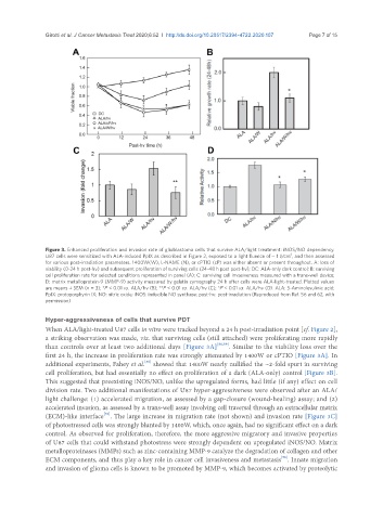
Figure 3. Enhanced proliferation and invasion rate of glioblastoma cells that survive ALA/light treatment: iNOS/NO dependency.
2
U87 cells were sensitized with ALA-induced PpIX as described in Figure 2, exposed to a light fluence of ~ 1 J/cm , and then assessed
for various post-irradiation parameters. 1400W(W), L-NAME (N), or cPTIO (cP) was either absent or present throughout. A: loss of
viability (0-24 h post-hν) and subsequent proliferation of surviving cells (24-48 h post post-hν); DC: ALA-only dark control; B: surviving
cell proliferation rate for selected conditions represented in panel (A); C: surviving cell invasiveness measured with a trans-well device;
D: matrix metalloprotein-9 (MMP-9) activity measured by gelatin zymography 24 h after cells were ALA-light-treated. Plotted values
are means ± SEM (n = 3); *P < 0.01 vs. ALA/hν (B); **P < 0.01 vs. ALA/hν (C); *P < 0.01 vs. ALA/hν (D). ALA: 5-Aminolevulinic acid;
PpIX: protoporphyrin IX; NO: nitric oxide; iNOS: inducible NO synthase; post-hν: post-irradiation (Reproduced from Ref. 56 and 62, with
permission)
Hyper-aggressiveness of cells that survive PDT
When ALA/light-treated U87 cells in vitro were tracked beyond a 24 h post-irradiation point [cf. Figure 2],
a striking observation was made, viz. that surviving cells (still attached) were proliferating more rapidly
than controls over at least two additional days [Figure 3A] [56,59] . Similar to the viability loss over the
first 24 h, the increase in proliferation rate was strongly attenuated by 1400W or cPTIO [Figure 3A]. In
[56]
additional experiments, Fahey et al. showed that 1400W nearly nullified the ~2-fold spurt in surviving
cell proliferation, but had essentially no effect on proliferation of a dark (ALA-only) control [Figure 3B].
This suggested that preexisting iNOS/NO, unlike the upregulated forms, had little (if any) effect on cell
division rate. Two additional manifestations of U87 hyper-aggressiveness were observed after an ALA/
light challenge: (1) accelerated migration, as assessed by a gap-closure (wound-healing) assay; and (2)
accelerated invasion, as assessed by a trans-well assay involving cell traversal through an extracellular matrix
[56]
(ECM)-like interface . The large increase in migration rate (not shown) and invasion rate [Figure 3C]
of photostressed cells was strongly blunted by 1400W, which, once again, had no significant effect on a dark
control. As observed for proliferation, therefore, the more aggressive migratory and invasive properties
of U87 cells that could withstand photostress were strongly dependent on upregulated iNOS/NO. Matrix
metalloproteinases (MMPs) such as zinc-containing MMP-9 catalyze the degradation of collagen and other
[56]
ECM components, and thus play a key role in cancer cell invasiveness and metastasis . Innate migration
and invasion of glioma cells is known to be promoted by MMP-9, which becomes activated by proteolytic