
Page 60 - Read Online
P. 60
Page 4 of 9 Ballarò et al. J Cancer Metastasis Treat 2019;5:61 I http://dx.doi.org/10.20517/2394-4722.2019.003
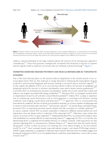
Figure 1. Exercise mimetics may positively affect cachexia progression and low exercise performance. In cancer patients, tumor growth
and chemotherapy may induce anemia, cardiac dysfunction and mitochondrial alterations, leading to loss of muscle mass and reduced
exercise performance. Exercise-like drugs may relieve muscle wasting and/or support cancer patients practicing exercise
addition, exercise performed in late-stage cachexia reduces the survival of C26-bearing mice exposed to
[21]
chemotherapy . Given these premises, strategies that circumvent the limitations of exercise or improve
[44]
exercise capacity could be useful for cancer hosts who are intolerant to physical training [Figure 1].
TARGETING EXERCISE-INDUCED PATHWAYS AND MUSCLE METABOLISM AS THERAPEUTIC
STRATEGY
One of the most relevant player in the exercise-induced adaptations in the skeletal muscle is the co-
transcription factor PGC-1α, that responds to energy demand by enhancing the transcription of genes
involved in mitochondrial biogenesis and turnover, leading to increased muscle oxidative capacity [44,45] .
In this regard, the ablation of PGC-1α in mice has been shown to blunt the increase of autophagy and
mitophagy induced by exercise, to decrease mitochondrial mass and to impair exercise performance [45,46] .
Conversely, PGC-1α overexpression increases mitochondrial content, fast-to-slow muscle fiber switch and
induces a set of genes associated with energy metabolism [47,48] . Moreover, PGC-1α transgenic animals show
[49]
an improvement of satellite cell activation and proliferative potential . Indeed, PGC-1α plays an important
role in modulating muscle homeostasis and its downregulation is correlated with different wasting
conditions, such as aging, muscle disuse and denervation [50-52] . In aged mice, PGC-1α overexpression has
been shown to counteract the loss of muscle mitochondrial enzymes, to increase markers of autophagy and
[47]
[53]
to preserve both neuromuscular junctions and exercise capacity . In muscle disuse and denervation,
increased PGC-1α expression prevents muscle atrophy and the activation of catabolic pathways [50,52] .
In cancer cachexia, muscle PGC-1α protein expression is reduced in different animal models and such
[54]
impairment does not always reflect a reduction of the transcript level . Otherwise, some studies also
[54]
reports an increase of PGC-1 α gene expression . Independently from PGC-1α, muscle mitochondrial mass
[10]
and metabolism are severely affected in tumor-bearing animals , suggesting that PGC-1α is not the only
determining factor in impaired mitochondrial function. In cachectic animals, conflicting results are also
reported regarding PGC-1α overexpression. Whereas Wang et al. demonstrate that the muscle-specific
[55]
upregulation of PGC-1α does not protect LLC-bearing mice from muscle atrophy in the same animal
model Pin et al. show increased muscle mass and reduced atrogin-1 and muscle RING-finger protein-1
[35]
(MuRF-1) gene expression as compared to wild-type tumor bearers. However, both the studies suggest that
forcing the expression of PGC-1α in the skeletal muscle could have some limitations, such as the increase
in tumor mass [35,55] .