
Page 99 - Read Online
P. 99
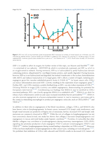
Page 10 of 18                        Wallace et al. J Cancer Metastasis Treat 2019;5:9  I  http://dx.doi.org/10.20517/2394-4722.2019.01


               Figure 1. BVD and LVD during mammary gland development. BVD measured as number of blood vessels per millimeter and LVD
               measured as number of lyve-1 positive vessels per millimeter in nulliparous (N), pregnant (P), lactating (L), involuting (days 2-10) and
                                                           [16]
               regressed (R), mammary glands (data adapted from Lyons et al.  and Ramirez et al. [144] ). BVD: Blood vessel density; LVD: lymphatic
               vessel density

               (HIF-1) is unable to affect its targets; for further review of this topic, see Masson and Ratcliffe [150] . HIF-
               1 is comprised of two subunits - ARNT/HIF-1b, which is constitutively expressed, and HIF-1α, which is
               an oxygen-sensitive subunit. During normoxia, HIF-1α is hydroxylated by prolyl hydroxylase domain
               containing proteins, ubiquitinated by von-Hippel-Lindau protein, and rapidly degraded. During hypoxia,
               however, HIF-1α is not hydroxylated and degraded, but instead, translocates to the nucleus, heterodimerizes
               with ARNT, and induces transcription of its target genes. HIF-1 affects a variety of targets, including pro-
               angiogenic genes like vascular endothelial growth factor-A (VEGF-A) [151] . In breast cancer cells, COX-2
                                                                                                       [48]
               can induce inflammation-associated HIF-1 activity, resulting in the expression of pro-angiogenic genes .
                                                                                                       [152]
               Further, HIF-1 and COX-2 maintain a positive feedback loop, as HIF-1 can also induce expression of COX-2 .
               Utilizing NSAIDs to target COX-2 activity can inhibit angiogenesis, demonstrating the potential for
               therapeutic intervention [152-154] . Corroborating our findings that SEMA7A may be involved in COX-2
               associated pathways, HIF-1 can directly upregulate SEMA7A in endothelial cells [155] . SEMA7A can elicit the
               release of pro-inflammatory cytokines and cause increased endothelial barrier permeability [156,157] . SEMA7A
               can also promote angiogenesis in a hypoxia-independent manner in murine mammary carcinoma and in
               the cornea by stimulating macrophages to produce pro-angiogenic molecules, such as CXCL2/MIP-2 [158]  and
               VEGF-A [159] .

               In addition to their roles in angiogenesis of the blood vasculature, collagen, COX-2, and SEMA7A also
               have known roles in lymphangiogenesis. In breast cancer, increased LVD, lymph node involvement, and
               lymph vessel invasion are predictive of higher risk of metastasis; further, increased LVD and lymph node
                                                     [16]
               metastasis are commonly observed in PPBC . While the role of collagen in lymphangiogenesis has not
               been extensively characterized, one study has shown that collagen I increases lymphangiogenesis and
               angiogenesis in mouse embryoid bodies under hypoxic conditions [160] . Therefore, it is plausible that other
               fibrillar collagens may contribute to lymphangiogenesis during mammary tumorigenesis. COX-2/PGE2
               signaling also promotes production of pro-angiogenic VEGF-A and pro-lymphangiogenic VEGF-C and
                                                                                              [162]
               VEGF-D  [161] ; further, lymphangiogenesis during wound healing is dependent on COX-2 activity . Moreover,
               COX-2 has been implicated in lymphangiogenesis in other cancer types, including cervical and gastric [163,164] .
               We published that inhibition of COX-2 with celecoxib and NSAIDs results in decreased LVD, tumor cell