
Page 250 - Read Online
P. 250
Page 10 of 18 D'Angelo et al. J Cancer Metastasis Treat 2019;5:30 I http://dx.doi.org/10.20517/2394-4722.2018.86
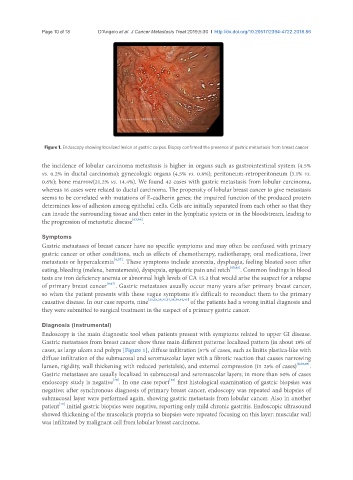
Figure 1. Endoscopy showing localized lesion at gastric corpus. Biopsy confirmed the presence of gastric metastasis from breast cancer
the incidence of lobular carcinoma metastasis is higher in organs such as gastrointestinal system (4.5%
vs. 0.2% in ductal carcinoma); gynecologic organs (4.5% vs. 0.8%); peritoneum-retroperitoneum (3.1% vs.
0.6%); bone marrow(21.2% vs. 14.4%). We found 42 cases with gastric metastasis from lobular carcinoma,
whereas 16 cases were related to ductal carcinoma. The propensity of lobular breast cancer to give metastasis
seems to be correlated with mutations of E-cadherin genes; the impaired function of the produced protein
determines loss of adhesion among epithelial cells. Cells are initially separated from each other so that they
can invade the surrounding tissue and then enter in the lymphatic system or in the bloodstream, leading to
the progression of metastatic disease [63,64] .
Symptoms
Gastric metastases of breast cancer have no specific symptoms and may often be confused with primary
gastric cancer or other conditions, such as effects of chemotherapy, radiotherapy, oral medications, liver
metastasis or hypercalcemia [4,57] . These symptoms include anorexia, dysphagia, feeling bloated soon after
eating, bleeding (melena, hematemesis), dyspepsia, epigastric pain and retch [65,66] . Common findings in blood
tests are iron deficiency anemia or abnormal high levels of CA 15.3 that would arise the suspect for a relapse
of primary breast cancer [9,67] . Gastric metastases usually occur many years after primary breast cancer,
so when the patient presents with these vague symptoms it’s difficult to reconduct them to the primary
causative disease. In our case reports, nine [23,28,29,35,37,38,39,43,45] of the patients had a wrong initial diagnosis and
they were submitted to surgical treatment in the suspect of a primary gastric cancer.
Diagnosis (instrumental)
Endoscopy is the main diagnostic tool when patients present with symptoms related to upper GI disease.
Gastric metastases from breast cancer show three main different patterns: localized pattern (in about 18% of
cases, as large ulcers and polyps [Figure 1], diffuse infiltration (57% of cases, such as linitis plastica-like with
diffuse infiltration of the submucosal and seromuscolar layer with a fibrotic reaction that causes narrowing
lumen, rigidity, wall thickening with reduced peristalsis), and external compression (in 25% of cases) [4,68,69] .
Gastric metastases are usually localized in submucosal and seromuscolar layers; in more than 50% of cases
[70]
[18]
endoscopy study is negative . In one case report first histological examination of gastric biopsies was
negative; after synchronous diagnosis of primary breast cancer, endoscopy was repeated and biopsies of
submucosal layer were performed again, showing gastric metastasis from lobular cancer. Also in another
[19]
patient initial gastric biopsies were negative, reporting only mild chronic gastritis. Endoscopic ultrasound
showed thickening of the muscolaris propria so biopsies were repeated focusing on this layer: muscular wall
was infiltrated by malignant cell from lobular breast carcinoma.