
Page 200 - Read Online
P. 200
Page 10 of 18 Borniger. J Cancer Metastasis Treat 2019;5:23 I http://dx.doi.org/10.20517/2394-4722.2018.107
[83]
treatments . Additionally, due to its low toxicity and high tolerability, it may be useful as a powerful and
inexpensive adjunct therapy.
Midbrain reward system
The midbrain ventral tegmental area (VTA) and neighboring substantia nigra are the primary source of all
dopamine (DA) within the brain. Known for its important role in reward and motivational processing (i.e.,
calculating reward-prediction errors), the VTA has recently become a target for modulating cancer. Elevated
concentrations of dopamine are associated with blunted tumor growth, reduced angiogenesis, and lower
[84]
metastatic capacity of cancer in rats . In general, dopamine seems to inhibit cancer growth, while serotonin
[85]
facilitates it . The mechanisms underlying this phenomenon are unclear, although research has started to
make headway in this area. In recent years, the VTA has been linked to the modulation of both innate and
[86]
adaptive immunity . Using designer receptors exclusively activated by designer drugs (DREADDs), Rolls
and colleagues demonstrated that activation of VTA-DA neurons promotes monocyte/macrophage expansion
and innate immune responses to E. coli infection. Activation of these neurons further increased the number
of circulating B-cells, subsequent IgM and IgG titers in response to E. coli, and interferon-g production by
T-cells, suggesting enhanced adaptive immunity.
[87]
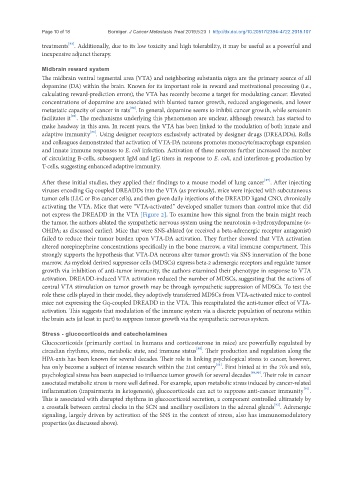
After these initial studies, they applied their findings to a mouse model of lung cancer . After injecting
viruses encoding Gq-coupled DREADDs into the VTA (as previously), mice were injected with subcutaneous
tumor cells (LLC or B16 cancer cells), and then given daily injections of the DREADD ligand CNO, chronically
activating the VTA. Mice that were “VTA-activated” developed smaller tumors than control mice that did
not express the DREADD in the VTA [Figure 2]. To examine how this signal from the brain might reach
the tumor, the authors ablated the sympathetic nervous system using the neurotoxin 6-hydroxydopamine (6-
OHDA; as discussed earlier). Mice that were SNS-ablated (or received a beta-adrenergic receptor antagonist)
failed to reduce their tumor burden upon VTA-DA activation. They further showed that VTA activation
altered norepinephrine concentrations specifically in the bone marrow, a vital immune compartment. This
strongly supports the hypothesis that VTA-DA neurons alter tumor growth via SNS innervation of the bone
marrow. As myeloid derived suppressor cells (MDSCs) express beta-2 adrenergic receptors and regulate tumor
growth via inhibition of anti-tumor immunity, the authors examined their phenotype in response to VTA
activation. DREADD-induced VTA activation reduced the number of MDSCs, suggesting that the actions of
central VTA stimulation on tumor growth may be through sympathetic suppression of MDSCs. To test the
role these cells played in their model, they adoptively transferred MDSCs from VTA-activated mice to control
mice not expressing the Gq-coupled DREADD in the VTA. This recapitulated the anti-tumor effect of VTA-
activation. This suggests that modulation of the immune system via a discrete population of neurons within
the brain acts (at least in part) to suppress tumor growth via the sympathetic nervous system.
Stress - glucocorticoids and catecholamines
Glucocorticoids (primarily cortisol in humans and corticosterone in mice) are powerfully regulated by
[88]
circadian rhythms, stress, metabolic state, and immune status . Their production and regulation along the
HPA-axis has been known for several decades. Their role in linking psychological stress to cancer, however,
[11]
has only become a subject of intense research within the 21st century . First hinted at in the 70’s and 80’s,
psychological stress has been suspected to influence tumor growth for several decades [89,90] . Their role in cancer
associated metabolic stress is more well defined. For example, upon metabolic stress induced by cancer-related
[91]
inflammation (impairments in ketogenesis), glucocorticoids can act to suppress anti-cancer immunity .
This is associated with disrupted rhythms in glucocorticoid secretion, a component controlled ultimately by
[92]
a crosstalk between central clocks in the SCN and ancillary oscillators in the adrenal glands . Adrenergic
signaling, largely driven by activation of the SNS in the context of stress, also has immunomodulatory
properties (as discussed above).