
Page 133 - Read Online
P. 133
Grelet et al. J Cancer Metastasis Treat 2019;5:16 I http://dx.doi.org/10.20517/2394-4722.2018.85 Page 3 of 10
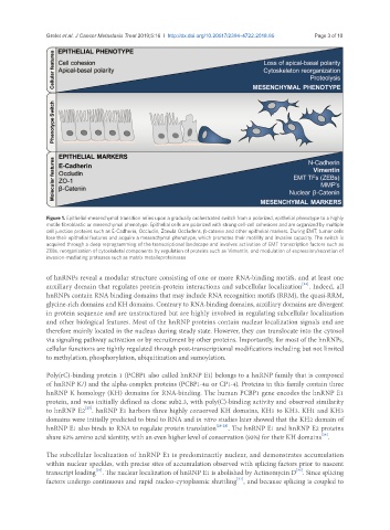
Figure 1. Epithelial-mesenchymal transition relies upon a gradually orchestrated switch from a polarized, epithelial phenotype to a highly
motile fibroblastic or mesenchymal phenotype. Epithelial cells are polarized with strong cell-cell cohesions and are organized by multiple
cell junction proteins such as E-Cadherin, Occludin, Zonula Occludens, β-catenin and other epithelial markers. During EMT, tumor cells
lose their epithelial features and acquire a mesenchymal phenotype, which promotes their motility and invasive capacity. The switch is
acquired through a deep reprogramming of the transcriptional landscape and involves activation of EMT transcription factors such as
ZEBs, reorganization of cytoskeletal components by regulation of proteins such as Vimentin, and modulation of expression/secretion of
invasion-mediating proteases such as matrix metalloproteinases
of hnRNPs reveal a modular structure consisting of one or more RNA-binding motifs, and at least one
[24]
auxiliary domain that regulates protein-protein interactions and subcellular localization . Indeed, all
hnRNPs contain RNA binding domains that may include RNA recognition motifs (RRM), the quasi-RRM,
glycine-rich domains and KH domains. Contrary to RNA-binding domains, auxiliary domains are divergent
in protein sequence and are unstructured but are highly involved in regulating subcellular localization
and other biological features. Most of the hnRNP proteins contain nuclear localization signals and are
therefore mainly located in the nucleus during steady state. However, they can translocate into the cytosol
via signaling pathway activation or by recruitment by other proteins. Importantly, for most of the hnRNPs,
cellular functions are tightly regulated through post-transcriptional modifications including but not limited
to methylation, phosphorylation, ubiquitination and sumoylation.
Poly(rC)-binding protein 1 (PCBP1 also called hnRNP E1) belongs to a hnRNP family that is composed
of hnRNP K/J and the alpha-complex proteins (PCBP1-4α or CP1-4). Proteins in this family contain three
hnRNP K homology (KH) domains for RNA-binding. The human PCBP1 gene encodes the hnRNP E1
protein, and was initially defined as clone sub2.3, with poly(C)-binding activity and observed similarity
[25]
to hnRNP E2 . hnRNP E1 harbors three highly conserved KH domains, KH1 to KH3. KH1 and KH3
domains were initially predicted to bind to RNA and in vitro studies later showed that the KH2 domain of
hnRNP E1 also binds to RNA to regulate protein translation [26-28] . The hnRNP E1 and hnRNP E2 proteins
[24]
share 82% amino acid identity, with an even higher level of conservation (93%) for their KH domains .
The subcellular localization of hnRNP E1 is predominantly nuclear, and demonstrates accumulation
within nuclear speckles, with precise sites of accumulation observed with splicing factors prior to nascent
[30]
[29]
transcript loading . The nuclear localization of hnRNP E1 is abolished by Actinomycin D . Since splicing
[31]
factors undergo continuous and rapid nucleo-cytoplasmic shuttling , and because splicing is coupled to