
Page 36 - Read Online
P. 36
Page 14 of 25 Kondapuram et al. J Cancer Metastasis Treat 2019;5:32 I http://dx.doi.org/10.20517/2394-4722.2018.105
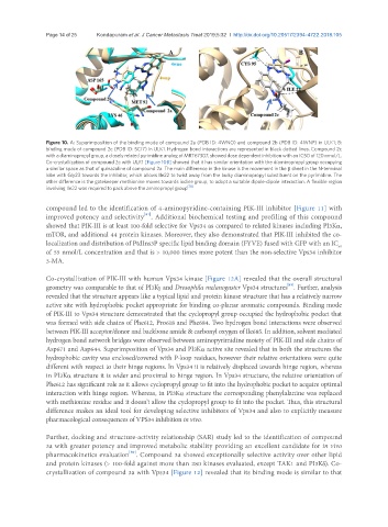
Figure 10. A: Superimposition of the binding mode of compound 2a (PDB ID: 4WNO) and compound 2b (PDB ID: 4WNP) in ULK1; B:
binding mode of compound 2c (PDB ID: 5CI7) in ULK1. Hydrogen bond interactions are represented in black dotted lines. Compound 2c
with a diaminopropyl group, a closely related pyrimidine analog of MRT67307, showed dose dependent inhibition with an IC50 of 120 nmol/L.
Co-crystallization of compound 2c with ULK1 [Figure 10B] showed that it has similar orientation with the diaminopropyl group occupying
a similar space as that of quinazoline of compound 2a. The main difference in the kinase is the movement in the β sheet in the N-terminal
lobe with Gly23 towards the inhibitor, which allows Ile22 to twist away from the bulky diaminopropyl substituent on the pyrimidine. The
other difference is the gatekeeper methionine moves towards iodine group, to adopt a suitable dipole-dipole interaction. A flexible region
involving Ile22 was required to pack above the aminopropyl group [78]
compound led to the identification of 4-aminopyridine-containing PIK-III inhibitor [Figure 11] with
[81]
improved potency and selectivity . Additional biochemical testing and profiling of this compound
showed that PIK-III is at least 100-fold selective for Vps34 as compared to related kinases including PI3Kα,
mTOR, and additional 44 protein kinases. Moreover, they also demonstrated that PIK-III inhibited the co-
localization and distribution of PtdIns3P specific lipid binding domain (FYVE) fused with GFP with an IC
50
of 55 nmol/L concentration and that is > 10,000 times more potent than the non-selective Vps34 inhibitor
3-MA.
Co-crystallization of PIK-III with human Vps34 kinase [Figure 12A] revealed that the overall structural
[81]
geometry was comparable to that of PI3Kγ and Drosophila melanogaster Vps34 structures . Further, analysis
revealed that the structure appears like a typical lipid and protein kinase structure that has a relatively narrow
active site with hydrophobic pocket appropriate for binding co-planar aromatic compounds. Binding mode
of PIK-III to Vps34 structure demonstrated that the cyclopropyl group occupied the hydrophobic pocket that
was formed with side chains of Phe612, Pro618 and Phe684. Two hydrogen bond interactions were observed
between PIK-III acceptor/donor and backbone amide & carbonyl oxygen of Ile685. In addition, solvent mediated
hydrogen bond network bridges were observed between aminopyrimidine moiety of PIK-III and side chains of
Asp671 and Asp644. Superimposition of Vps34 and PI3Kα active site revealed that in both the structures the
hydrophobic cavity was enclosed/covered with P-loop residues, however their relative orientations were quite
different with respect to their hinge regions. In Vps34 it is relatively displaced towards hinge region, whereas
in PI3Kα structure it is wider and proximal to hinge region. In Vps34 structure, the relative orientation of
Phe612 has significant role as it allows cyclopropyl group to fit into the hydrophobic pocket to acquire optimal
interaction with hinge region. Whereas, in PI3Kα structure the corresponding phenylalanine was replaced
with methionine residue and it doesn’t allow the cyclopropyl group to fit into the pocket. Thus, this structural
difference makes an ideal tool for developing selective inhibitors of Vps34 and also to explicitly measure
pharmacological consequences of VPS34 inhibition in vivo.
Further, docking and structure-activity relationship (SAR) study led to the identification of compound
3a with greater potency and improved metabolic stability providing an excellent candidate for in vivo
[80]
pharmacokinetics evaluation . Compound 3a showed exceptionally selective activity over other lipid
and protein kinases (> 100-fold against more than 280 kinases evaluated, except TAK1 and PI3Kδ). Co-
crystallization of compound 3a with Vps34 [Figure 12] revealed that its binding mode is similar to that