
Page 54 - Read Online
P. 54
Jusino et al. J Cancer Metastasis Treat 2018;4:43 I http://dx.doi.org/10.20517/2394-4722.2018.24 Page 7 of 20
Centrosome disengagement
Separase
and Plk1
MC
DC
Centrosome disjunction, Procentriole assembly
separation and maturation centrosome duplication
Cdk1, Cdk2, SAS5/
Nek2, Ana2/STIL,
Aurora A SAS-6,
and Plk1 ZYG1/Plk4
Procentriole
Elongation
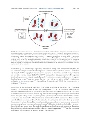
Figure 1. The centrosome duplication cycle. The mother centriole (MC) is depicted with blue triangles that represent the distal and
sub-distal appendages to differentiate it from the daughter centriole (DC). In the G1 phase, the two centrioles are connected by a
proteinaceous linker. The G1/S transition phase is characterized by the procentriole assembly, and some of the key proteins involved in
this process are mentioned. In this stage, the DC starts to acquire the appendages that the MC has. During the S phase, the microtubules
are synthesized, and rearrangement will occur to fully generate the procentriole. Till the G2 phase, the proteinaceous linker is broken,
and the DC already has the distal and sub-distal appendages. This will convert DC into MC, and two pairs of centrioles will be formed. In
the G2/M transition phase centrosome disjunction, separation, and maturation take place. Some key regulators have been listed above.
During the M phase, the separated centrioles participate in bipolar spindle mitosis, and the centrosome cycle is completed when each
daughter cell inherits two centrioles
phosphorylating and inactivating c-Nap1 and β-catenin [123,124] . Lastly, from metaphase to anaphase, the
two centrosomes migrate to opposite cellular poles and form the mitotic spindle to which the kinetochore
will attach . Faithful segregation of chromosomes is ensured by the spindle assembly checkpoint (SAC)
[82]
and associated proteins such as BUB1B , MPS1 , among others. Other proteins that play important
[125]
[126]
functions in chromosome integrity include Bub1, which maintains sister chromatid cohesion through the
phosphorylation of SgoI ; another protein that plays a key role in this activity is PP2A, which ensures
[127]
localization of Sgo1 to centromeres . Aurora kinase B, survivin, and ICENP play important roles in
[128]
cytokinesis [Figure 1].
[129]
Deregulation of the centrosome duplication cycle results in centrosome aberrations and chromosome
instability that ultimately have an effect on tumorigenesis [87,88,130] . While centrosome aberrations are
traditionally associated with cancer, mutations in genes that codify for centrosome proteins are also known
to cause human diseases such as ciliopathies (e.g., autosomal recessive primary microcephaly, Bardet-Biedl
disease, polycystic kidney disease, and primary ciliary dyskinesia) . Centrosome aberrations are classified
[131]
as numerical and structural . Both aberrations co-occur in tumors [133,134] . Centrosome aberrations have
[132]
been identified in most cancer types . For example, pioneering studies from the Doxsey laboratory
[94]
demonstrated structural abnormalities in number, position, shape, and size of centrosomes in primary solid
tumors, including brain, breast, colon, lung, and prostate . Likewise, studies from the Salisbury laboratory
[92]
showed that breast cancer tissue displayed abnormal structural and numerical centrosome aberrations,
abnormal mitoses and chromosome instability relative to normal breast tissue [133,135,136] and that centrosome
amplification in breast cancers is indicative of tumor aggressiveness .
[137]