
Page 51 - Read Online
P. 51
Page 4 of 20 Jusino et al. J Cancer Metastasis Treat 2018;4:43 I http://dx.doi.org/10.20517/2394-4722.2018.24
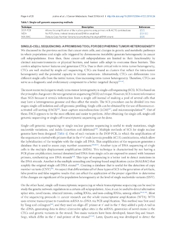
Table 1. Single-cell genomic sequencing methods
Technique Description References
DOP-PCR Allows the amplification of the nucleus genome using primers with ACTG combinations [52]
MDA No PCR phase; instead denaturalized DNA is amplified [53-55]
MALBAC Detects Copy Number Variants by amplifying the original DNA strand [53]
SINGLE-CELL SEQUENCING: A PROMISING TOOL FOR DECIPHERING TUMOR HETEROGENEITY
We discussed in the previous section that cancer stem cells, and changes in genetic and metabolic pathways
in whole populations and single cells triggered by chromosome instability generate heterogeneity in cancer
cell subpopulations. Even then, these cancer-cell subpopulations are limited in their functionality by
distinct microenvironments or physical barriers, and tumor cells adapt to overcome these barriers. This
confers adaptive tumor features and generates CTCs. Due to their critical role in intra-tumor heterogeneity,
CTCs are well studied by single-cell sequencing. CTCs are found as clusters that reflect the intra-tumor
heterogeneity and the potential capacity to initiate metastasis. Alternatively, CTCs can differentiate into
different single cells from the initial tumor, thus increasing intra-tumor heterogeneity. Therefore, CTCs can
serve as a diagnostic and evolutionary component to a better-targeted therapy [45-48] .
The most recent technique to study intra-tumor heterogeneity is single-cell sequencing (SCS). SCS is based on
the principles that govern the next generation sequencing (NGS) technique. However, SCS is more informative
than NGS because it reveals information from a single cell instead of making a pool of several cells that
may have a heterogeneous genome and thus affect the results. The SCS procedure can be divided into two
stages: single cell isolation and cell genomic profiling. Single cells can be obtained by the use of fluorescence-
activated cell sorting (FACS) , laser-capture microdissection (LCM) , and micromanipulation . Out of
[49]
[47]
[49]
these, FACS appears to be the most efficient and easier to perform. After obtaining the single cell, single-cell
genomic sequencing or single-cell transcriptomic sequencing can be done.
Single-cell genomic sequencing or single nuclear genome sequencing is useful to study mutations, single
nucleotide variations, and indels (insertion and deletions) . Multiple methods of SCS for single nuclear
[50]
genome have been designed [Table 1]. One of such variants is the DOP-PCR, in which the amplification of
the sequences is started with primers that in the 5’-3’ ends have six possible ACTG combinations, which allow
the hybridization of the template with the single cell DNA. This amplification of the sequences generates a
database that is used to assess copy number assessment [39,41,51] . Another type of DNA sequencing of single
cells is the multiple displacement amplification (MDA). This technique is characterized by not having a
PCR phase amplification; instead denaturalized DNA from single cells are exposed to anneal with hexamer
primers, synthesizing new DNA strands . This type of sequencing is a better tool to detect mutations in
[52]
the DNA strands. Another is the multiple annealing and looping-based amplification cycles (MALBAC) that
amplify the original single cell DNA strand . Creating a database that is useful for the detection of copy
[51]
number variants (CNV) . An aspect that differentiates all of these types of SCS is the generation of artifacts,
[53]
false positive and false negative results that can affect the application of the proper algorithm to determine
if the changes are significant of the population heterogeneity at the level of single nucleotide variants (SNV).
On the other hand, single-cell transcriptomic sequencing or whole transcriptome sequencing can be used to
study the genetic network regulation in a certain cell subpopulation. Also, it can be useful to detect alternative
splice sites, novel exons, retained introns, coding RNAs, and non-coding RNAs, among others [39,41,50] . Most
of the sequencing protocols in cancer research use the whole transcriptome amplification (WTA). WTA
uses reverse transcriptase to transform mRNA to cDNA via PCR amplification. This method was first used
by Tang and colleagues , and they used an oligo-dT primer at 5’ and in the 3’ they added a poly-A tail in
[56]
the cDNA, generating data to detect alternative splice sites in the mRNA, generation of novel exons in the
CTCs and genetic variants in the strand. Two main variants have been developed, Smart-Seq and Smart-
Seq2, which differ in the 5’ end primer of the strand [57,58] . Later, Quartz-seq was developed to detect the