
Page 29 - Read Online
P. 29
Page 6 of 9 Felton et al. J Cancer Metastasis Treat 2018;4:51 I http://dx.doi.org/10.20517/2394-4722.2018.39
A B E
C D F
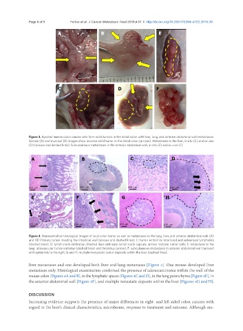
Figure 3. Injected human colon cancer cells form solid tumors in the distal colon with liver, lung, and anterior abdominal wall metastases.
Serosal (A) and mucosal (B) images show invasive solid tumor in the distal colon (arrows). Metastases in the liver, in situ (C) and ex vivo
(D) (arrows and dashed lines). Subcutaneous metastases in the anterior abdominal wall, in situ (E) and ex vivo (F).
A B C D
100 mm 200 mm 400 mm 100 mm
E F G H
100 mm 100 mm 100 mm 100 mm
Figure 4. Representative histological images of local colon tumor as well as metastases to the lung, liver, and anterior abdominal wall. (A)
and (B) Primary tumor invading the intestinal wall (arrows and dashed lines); C: tumor emboli in intramural and subserosal lymphatics
(dashed lines); D: lymph node infiltration. Dashed lines delineate lymph node capsule, arrows indicate tumor cells; E: metastasis to the
lung: intravascular tumor embolus (dashed lines) and thrombus (arrow); F: subcutaneous metastasis to anterior abdominal wall (arrows)
with epidermis to the right; G and H: multiple metastatic tumor deposits within the liver (dashed lines).
liver metastases and one developed both liver and lung metastases [Figure 3]. One mouse developed liver
metastases only. Histological examination confirmed the presence of adenocarcinoma within the wall of the
mouse colon [Figures 4A and B], in the lymphatic spaces [Figures 4C and D], in the lung parenchyma [Figure 4E], in
the anterior abdominal wall [Figure 4F], and multiple metastatic deposits within the liver [Figures 4G and H].
DISCUSSION
Increasing evidence supports the presence of major differences in right- and left-sided colon cancers with
regard to the host’s clinical characteristics, microbiome, response to treatment and outcome. Although mo-