
Page 28 - Read Online
P. 28
Felton et al. J Cancer Metastasis Treat 2018;4:51 I http://dx.doi.org/10.20517/2394-4722.2018.39 Page 5 of 9
A B C
Figure 1. Main steps in the surgical approach to injecting colon cancer cells in the murine distal colon. A: Isolation of the distal colon
(outlined) using moist sterile cotton tip applicators with retraction of the abdominal wall and evisceration of abdominal organs; B:
6
injection of 5 × 10 HT-29 human colon cancer cells into the wall of the distal colon (arrow) using a 27-guage needle; C: applying pressure
with a moist sterile cotton tip applicator at the injection site to prevent leakage and hemorrhage
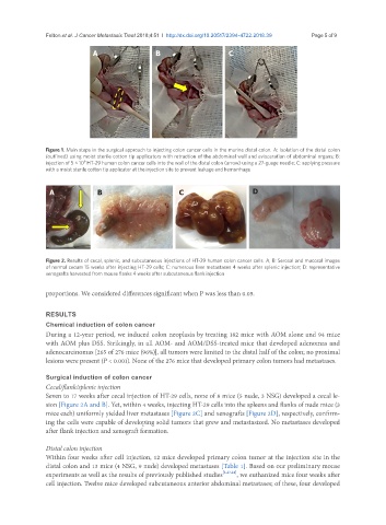
A B C D
Figure 2. Results of cecal, splenic, and subcutaneous injections of HT-29 human colon cancer cells. A, B: Serosal and mucosal images
of normal cecum 15 weeks after injecting HT-29 cells; C: numerous liver metastases 4 weeks after splenic injection; D: representative
xenografts harvested from mouse flanks 4 weeks after subcutaneous flank injection
proportions. We considered differences significant when P was less than 0.05.
RESULTS
Chemical induction of colon cancer
During a 12-year period, we induced colon neoplasia by treating 182 mice with AOM alone and 94 mice
with AOM plus DSS. Strikingly, in all AOM- and AOM/DSS-treated mice that developed adenomas and
adenocarcinomas [265 of 276 mice (96%)], all tumors were limited to the distal half of the colon; no proximal
lesions were present (P < 0.001). None of the 276 mice that developed primary colon tumors had metastases.
Surgical induction of colon cancer
Cecal/flank/splenic injection
Seven to 17 weeks after cecal injection of HT-29 cells, none of 8 mice (5 nude, 3 NSG) developed a cecal le-
sion [Figure 2A and B]. Yet, within 4 weeks, injecting HT-29 cells into the spleens and flanks of nude mice (3
mice each) uniformly yielded liver metastases [Figure 2C] and xenografts [Figure 2D], respectively, confirm-
ing the cells were capable of developing solid tumors that grew and metastasized. No metastases developed
after flank injection and xenograft formation.
Distal colon injection
Within four weeks after cell injection, 12 mice developed primary colon tumor at the injection site in the
distal colon and 13 mice (4 NSG, 9 nude) developed metastases [Table 1]. Based on our preliminary mouse
experiments as well as the results of previously published studies [1,23,24] , we euthanized mice four weeks after
cell injection. Twelve mice developed subcutaneous anterior abdominal metastases; of these, four developed