
Page 934 - Read Online
P. 934
Caron de Fromentel et al. Hepatoma Res 2020;6:80 I http://dx.doi.org/10.20517/2394-5079.2020.77 Page 3 of 18
A B
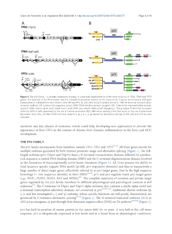
Figure 1. The p53 family - a complex expression strategy. A: schematic representation of the exon structure of TP53, TP63 and TP73
genes. The position of the P1 promoter and the internal P2 promoter relative to the exons in the 3 genes are indicated. p53 gene
transcription is initiated from two distinct sites (P1 and P1′); B: p53, p63 and p73 protein domains. TAD: N-terminal transactivation
domains (yellow); PR: proline-rich sequence (grey); DBD: DNA-binding domain (purple); OD: C-terminal oligomerization domain
(green); SAM: sterile alpha motif (dark blue); post-SAM: post-sterile alpha motif (burgundy). The principal N-terminal truncated
isoforms (ΔTA or ΔN), generated by the use of internal promoters (P2), alternative splicing of the first exons or the use of alternative
translation start sites, and the COOH-terminal variants a, β, g, d, e, z, generated by alternative splicing of p53, p63 and p73 are also
indicated
members and key players of stemness, which could help developing new approaches to prevent the
appearance of liver CSCs in the context of chronic liver diseases, inflammation in the liver, and HCC
development.
THE P53 FAMILY
The p53 family encompasses three members, namely TP53, TP63 and TP73 [18,19] . All three genes encode for
multiple isoforms generated by both internal promoter usage and alternative splicing [Figure 1]. The full-
length isoforms (p53, TAp63 and TAp73) share a N-terminal transactivation domain, followed by a proline-
rich sequence, a central DNA-binding domain (DBD) and the C-terminal oligomerization domain involved
in the formation of transcriptionally active homo-tetramers [Figure 1]. All three possess the ability to
bind sequence specific cognate DNA motifs (p53RE, p53 responsive elements) and thus to transactivate a
large number of direct target genes collectively referred to as p53-target genes. Due to the high sequence
homology (> 70% sequence identity) in their DBDs [18,19] , p73 and p63 regulate many p53-target genes
[20]
(e.g., WAF1, PUMA, NOXA, BAX and MDM2) . The complete repertoire of common and private target
genes regulated by the p53 family members in different physiological and pathological contexts is still
[20]
unknown . The C-terminus of TAp63 and TAp73 alpha isoforms also contains a sterile alpha motif and
a terminal transcription inhibitory domain, not conserved in p53 [18,19,21,22] . Additional shorter isoforms (β,
g, d, and less investigated e, z and h) isoforms, whose specific functions are still poorly characterized, are
generated by C-terminus alternative splicing [23,24] [Figure 1]. The N-terminal truncated isoforms (ΔTA or
ΔN) act as oncogenes, in part through their dominant negative effect (DNE) on TA isoforms [18,25,26] [Figure 1].
p53 has held its position of master protein in the cancer field for 40 years. A true hub in the cell stress
response, p53 is ubiquitously expressed at low levels and in a latent form in physiological conditions.