
Page 619 - Read Online
P. 619
Page 4 of 11 Ichida et al. Hepatoma Res 2020;6:54 I http://dx.doi.org/10.20517/2394-5079.2020.59
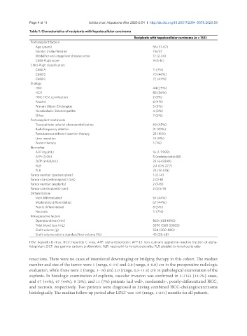
Table 1. Characteristics of recipients with hepatocellular carcinoma
Recipients with hepatocellular carcinoma (n = 153)
Pretransplant factors
Age (years) 56 (37-67)
Gender (male/female) 116/37
Model for end-stage liver disease score 12 (2-34)
Child-Pugh score 9 (5-14)
Child-Pugh classification
Child A 11 (7%)
Child B 70 (46%)
Child C 72 (47%)
Etiology
HBV 44 (29%)
HCV 85 (56%)
HBV, HCV, co-infection 2 (1%)
Alcohol 6 (4%)
Primary Biliary Cholangitis 5 (3%)
Nonalcoholic Steatohepatitis 4 (3%)
Other 7 (5%)
Pretransplant treatments
Transcatheter arterial chemoembolization 69 (45%)
Radiofrequency ablation 31 (20%)
Percutaneous ethanol injection therapy 23 (15%)
Liver resection 14 (9%)
Proton therapy 1 (1%)
Biomarker
AFP (ng/mL) 16 (1-11999)
AFP-L3 (%) 5 (undetectable-88)
DCP (mAU/mL) 33 (6-13248)
NLR 2.4 (0.5-27.7)
PLR 74 (18-578)
Tumor number (pretransplant) 1 (0-14)
Tumor size (pretransplant) (cm) 2 (0-8)
Tumor number (explants) 2 (1-19)
Tumor size (explants) (cm) 2 (0.5-11)
Differentiation
Well differentiated 67 (44%)
Moderately differentiated 67 (44%)
Poorly differentiated 8 (5%)
Necrosis 11 (7%)
Intraoperative factors
Operation time (min) 860 (601-1890)
Total blood loss (mL) 5390 (568-53835)
Graft volume (g) 554 (300-880)
Graft volume ratio to standard liver volume (%) 45 (28-68)
HBV: hepatitis B virus; HCV: hepatitis C virus; AFP: alpha-fetoprotein; AFP-L3: lens culinaris agglutinin-reactive fraction of alpha-
fetoprotein; DCP: des-gamma-carboxy prothrombin; NLR: neutrophil-to-lymphocyte ratio; PLR: platelet-to-lymphocyte ratio
resections. There were no cases of intentional downstaging or bridging therapy in this cohort. The median
number and size of the tumor were 1 (range, 0-14) and 2.0 (range, 0-8.0) cm in the preoperative radiologic
evaluation, while these were 2 (range, 1-19) and 2.0 (range, 0.5-11.0) cm in pathological examination of the
explants. In histologic examination of explants, vascular invasion was confirmed in 21/153 (13.7%) cases,
and 67 (44%), 67 (44%), 8 (5%), and 11 (7%) patients had well-, moderately-, poorly-differentiated HCC,
and necrosis, respectively. Two patients were diagnosed as having combined HCC-cholangiocarcinoma
histologically. The median follow-up period after LDLT was 139 (range, 1-231) months for all patients.