
Page 547 - Read Online
P. 547
Page 4 of 7 Boortalary et al. Hepatoma Res 2020;6:48 I http://dx.doi.org/10.20517/2394-5079.2020.38
A B
C D
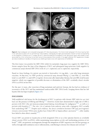
Figure 3. Chest radiograph and computed tomography (CT) showing empyema. The frontal chest radiograph (A) shows layering fluid
in the right pleural space with an air-fluid level more medially, overlapping the right paramediastinal shadow (arrow) corresponding
to the empyema. Enhanced axial CT images (B and C) show loculated fluid (asterisks) in the right pleural space with pockets of gas
surrounded by a thick, enhancing rim (arrows) typical of an empyema. More caudally in the upper abdomen (D), hepatectomy changes
are demonstrated post-resection of the previously noted large hepatic mass with regeneration of the left lobe
The liver tumor was positive for HBV DNA while the metastatic lung mass was negative for HBV DNA.
Serum samples from the time of her diagnosis of HCC and subsequent lung metastasis (both negative by
commercial assay) showed HBV DNA levels of 3,271 copies/mL and 52 copies/mL respectively.
Based on these findings, the patient was started on lamivudine 150 mg daily, 1 year after lung metastasis
resection. At that time, her HBV profile by commercial assay showed HBsAg (-), Anti-HBs (+), Anti-HBc
total (+), anti-HAV (+), and AFP 2.8 ng/mL. Seven years after lamivudine therapy, her anti-HBc total became
negative, which was suggestive of possible decrease or elimination of the HBV covalently closed circular
DNA (cccDNA) in her liver.
For the past 12 years, after resection of lung metastasis and antiviral therapy, she has had no evidence of
recurrence of the HCC and has maintained undetectable HBV DNA levels. Imaging shows that her left
hepatic lobe has hypertrophied [Figure 3D].
DISCUSSION
Well-established risk factors for the development of HCC in patients with chronic HBV infection are viral
load and the presence of HBeAg and HBsAg [8-10] . However, studies have demonstrated a high rate of OBI in
patients with HCC who are immunocompromised during chemotherapy for malignancy [11,12] , as well as in
[13]
patients with hepatitis C . The 38%-73% of patients from endemic areas with cryptogenic HCC actually
have underlying OBI [13-15] . Despite such evidence, the direct correlation between OBI and carcinogenesis
remains controversial. While some studies have linked OBI to hepatocellular carcinoma [16,17] , other studies
have failed to show direct causality [15,18] .
Occult HBV can persist in hepatocytes as both integrated DNA or as a free episome known as covalently
closed circular DNA (cccDNA), while maintaining transcription activity and synthesizing proteins at low
[15]
levels . HBV can promote carcinogenesis through the integration of HBV sequences into the host genome, as
[19]
well as through mild continuous micro-inflammation, contributing to chronic liver disease and cirrhosis .