
Page 545 - Read Online
P. 545
Page 2 of 7 Boortalary et al. Hepatoma Res 2020;6:48 I http://dx.doi.org/10.20517/2394-5079.2020.38
A B
C D
* *
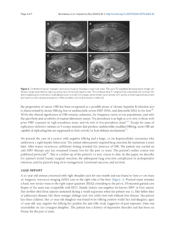
Figure 1. Contrast-enhanced magnetic resonance imaging showing a large liver mass. The axial T2-weighted fat-suppressed image (A)
shows a large hyperintense mass replacing most of the right hepatic lobe. The corresponding T1-weighted fat-suppressed pre-contrast (B),
arterial phase post-contrast (C) and delayed post-contrast (D) images demonstrate hypointensity with patchy arterial hyperenhancement
and washout with capsule appearance of the periphery and central necrosis (asterisk)
the progression of cancer. OBI has been recognized as a possible phase of chronic hepatitis B infection and
[2]
is characterized by absent HBsAg, low or undetectable serum HBV-DNA, and detectable DNA in the liver .
While the clinical significance of OBI remains unknown, the frequency varies across populations, and with
the specificity and sensitivity of routine laboratory assays. The prevalence is as high as 41%-90% in those with
[3,4]
prior HBV exposure in high-prevalence areas, and 5%-20% in low-prevalence areas . Except for cases of
replication-defective variants or S escape mutants that produce undetectable modified HBsAg, most OBI are
capable of replicating but are suppressed in their activity by host defense mechanisms .
[5]
We present the case of a patient with negative HBsAg and a large, 10 cm hepatocellular carcinoma who
underwent a right hepatic lobectomy. The patient subsequently required lung resection for metastasis 4 years
later. After tumor recurrence, additional testing revealed the presence of OBI. The patient was started on
anti-HBV therapy and has remained disease free for the past 16 years. The patient’s earlier course was
[6]
published previously . This is a follow up of the patient’s 16-year course to date. In this paper, we describe
the patient’s initial hepatic surgical resection, the subsequent lung resection complicated by postoperative
infection, and the patient’s long-term management, functional outcome, and survival.
CASE REPORT
A 64-year-old woman presented with right shoulder pain for one month and was found to have a 9 cm mass
on magnetic resonance imaging (MRI) scan in the right lobe of the liver [Figure 1]. Physical exam revealed
a hard, non-tender mass in the right upper quadrant (RUQ) extending to the pelvis. Ultrasound-guided core
biopsy of the mass was compatible with HCC. Family history was negative for known HBV or liver cancer.
Her mother died from injuries sustained during a bomb explosion when the patient was 14. Her father died
of pulmonary disease. Her three younger siblings (60F, 56F, 52M) were well without liver disease. The patient
has three children. Her 43-year-old daughter was found to be HBsAg positive whilst her 2nd daughter, aged
37-year-old, was negative for HBsAg but positive for anti-HBc total, suggestive of past exposure. Data was
unavailable on her youngest daughter. The patient has a history of depressive disorder and has been on
Prozac for the past 12 years.