
Page 47 - Read Online
P. 47
Page 4 of 13 Spieler et al. Hepatoma Res 2019;5:4 I http://dx.doi.org/10.20517/2394-5079.2018.77
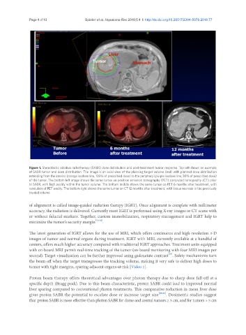
Figure 1. Stereotactic ablative radiotherapy (SABR) dose distribution and post-treatment tumor response. Top left shows an example
of SABR tumor and dose distribution. The image is an axial view of the planning target volume (red) with planned dose distribution
extending from the interior (orange isodose line, 105% of prescribed dose) to the periphery (purple isodose line, 30% of prescribed dose)
of the tumor. The bottom left image shows the same tumor on positron emission tomography (PET) computed tomography (CT) prior
to SABR, with high avidity within the tumor volume. The bottom middle shows the same tumor on PET 6 months after treatment, with
resolution of PET avidity. The bottom right shows the same tumor on CT 12 months after treatment, with tissue necrosis in the previously
treated volume
of alignment is called image-guided radiation therapy (IGRT). Once alignment is complete with millimeter
accuracy, the radiation is delivered. Currently most IGRT is performed using X-ray images or CT scans with
or without fiducial markers. Together, custom immobilization, respiratory management and IGRT help to
minimize the tumor’s security margin [16-18] .
The latest generation of IGRT allows for the use of MRI, which offers continuous and high-resolution 3-D
images of tumor and normal organs during treatment. IGRT with MRI, currently available at a handful of
centers, offers much higher accuracy compared with traditional IGRT approaches. Treatment units equipped
with on-board MRI permit real-time tracking of the tumor (on-board monitoring with four MRI images per
[19]
second). Target visualization can be further improved using gadoxetate contrast . Safety mechanisms turn
the beam off when the target transgresses the tracking volume, making it very safe to deliver high doses to
tumor with tight margins, sparing adjacent organs-at-risk [Video 1].
Proton beam therapy offers theoretical advantages over photon therapy due to sharp dose fall-off at a
specific depth (Bragg peak). Due to this beam characteristic, proton SABR could lead to improved normal
liver sparing compared to conventional photon treatments. This comparative reduction in mean liver dose
gives proton SABR the potential to escalate dose or increase target size [20,21] . Dosimetric studies suggest
that proton SABR is more effective than photon SABR for dome and central tumors ≥ 3 cm, and for tumors > 5 cm