
Page 645 - Read Online
P. 645
Page 6 of 12 Loria et al. Hepatoma Res 2018;4:59 I http://dx.doi.org/10.20517/2394-5079.2018.75
Table 2. Development of hepatocellular carcinoma (HCC)
Large regenerative nodule
Low dysplastic nodule
High dysplastic nodule
Nodule of HCC
well differentiated - moderately differentiated - poorly differentiated
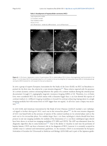
A B
C D
Figure 4. A: US shows a hypoechoic nodule; B: portal phase (32 s): arterial phase (23 s) shows a homogeneous isoenhancement of the
lesion; C: portal phase (52 s): the nodule is isoechoic with respect to the surrounding liver; D: late portal phase (280 s): the hepatocellular
carcinoma (HCC) is isoechoic with respect to the surrounding liver
In 2001 a group of experts European Association for the Study of the Liver (EASL) on HCC in Barcelona re-
[64]
ported, for the first time, the criteria for a non invasive diagnosis . These criteria required only the presence
of a certain dynamic contrast enhancing behavior: the uptake of a contrast medium during the arterial phase
documented through CT, angiography magnetic resonance imaging (MRI) or US. Therefore, in a cirrhotic
liver, were considered HCC the nodule lesions with a diameter bigger than 2 cm that showed this uptake of
contrast medium in 2 different imaging modalities or showed this contrast enhancing impregnation in a single
imaging modality but with serum levels of AFP bigger than 400 ng/mL. In all other cases a biopsy was neces-
sary [22,64] .
In 2005 EASL and American Association for the Study of Liver Diseases (AASLD) reached a new radiologi-
cal signal to further distinguish HCC: wash-out in the venous/late phase [5,22] . So the non invasive diagnosis
of HCC was based both on the presence of uptake of the contrast medium in the arterial phase and on the
wash-out in the venous/late phase. For nodules larger than 2 cm these radiological criteria should have been
present in just one imaging modality; for nodules of the dimensions of 1-2 cm these radiological signs should
have been shown in at least two imaging modalities (CT, MRI and CEUS). The AFP was eliminated from the
diagnostic algorithm due to some limitations [5,22] . Due to the ability to visualize in real time the perfusion of
hepatic lesions, CEUS can have a foremost role in the diagnosis of HCC; however it is currently accepted in
variable ways in national and international guidelines. At the moment, CEUS is recommended by European
Federation of Societies for Ultrasound in Medicine and Biology (EFSUMB) and is part of the Japanese guide-