
Page 643 - Read Online
P. 643
Page 4 of 12 Loria et al. Hepatoma Res 2018;4:59 I http://dx.doi.org/10.20517/2394-5079.2018.75
A B
C D
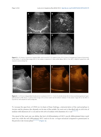
Figure 1. A: US shows a hypoechoic hepatocellular carcinoma (HCC); B: arterial phase (19 s) shows a homogeneous hyper-enhancement
of the lesion; C: portal phase image (82 s): the nodule is isoechoic; D: late portal phase (190 s): the HCC is slightly hypoechoic with
respect to surrounding liver
A B C D
Figure 2. A: US shows a hypoechoic hepatocellular carcinoma (HCC) (> 2 cm); B: arterial phase (26 s) shows an inhomogeneous hyper-
enhancement of the lesion; C: portal phase image (70 s) shows wash-out of contrast medium; D: late phase image (95 s): the HCC is
hypoechoic with respect to surrounding liver
To increase the specificity of CEUS on the basis of these findings, a demonstration of the washout-phase is
decisive and its presence also depends on the size of the nodule: the wash-out is described only in 20%-30% of
nodules with diameters of 1-2 cm and in 40%-60% of nodules with diameters of 2-3 cm [22,38,42-59] .
The speed of the wash-out can define the level of differentiation of HCC: poorly differentiated show rapid
wash-out, while the well differentiated HCC tends to be iso- or hypo-enhanced compared to parenchyma in
the portal or late venous phase [21,31,60-62] [Figure 4].