
Page 644 - Read Online
P. 644
Loria et al. Hepatoma Res 2018;4:59 I http://dx.doi.org/10.20517/2394-5079.2018.75 Page 5 of 12
Table 1. Typical enhancement of hepatocellular carcinoma in the arterial
phase based on the size of lesion
Size lesion (cm) rate of detection of the hyper-enhancement in lesion
< 1.0 cm 67%
1-2 cm 83%-88%
2-3 cm 92%-100%
A B
C D
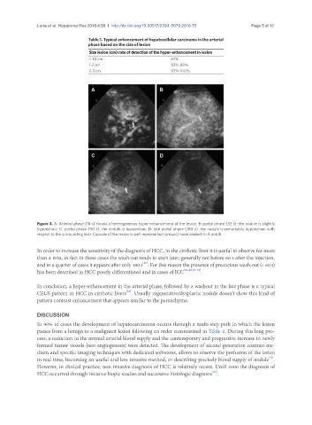
Figure 3. A: Arterial phase (18 s) shows a heterogeneous hyper-enhancement of the lesion; B: portal phase (32 s): the nodule is slightly
hypoechoic; C: portal phase (90 s): the nodule is hypoechoic; D: late portal phase (180 s): the nodule is remarkably hypoechoic with
respect to the surrounding liver. Capsule of the lesion is well represented (arrows) more evident in A and B
In order to increase the sensitivity of the diagnosis of HCC, in the cirrhotic liver it is useful to observe for more
than 4 min, in fact in these cases the wash-out tends to start later, generally not before 60 s after the injection,
[40]
and in a quarter of cases it appears after only 180 s . For this reason the presence of precocious wash-out (< 60 s)
has been described in HCC poorly differentiated and in cases of ICC [22,40,61-62] .
In conclusion, a hyper-enhancement in the arterial phase, followed by a washout in the late phase is a typical
[63]
CEUS pattern in HCC in cirrhotic livers . Usually regenerative/dysplastic nodule doesn’t show this kind of
pattern contrast enhancement that appears similar to the parenchyma.
DISCUSSION
In 90% of cases the development of hepatocarcinoma occurs through a multi-step path in which the lesion
passes from a benign to a malignant lesion following an order summarized in Table 2. During this long pro-
cess, a reduction in the normal arterial blood supply and the contemporary and progressive increase in newly
formed tumor vessels (neo-angiogenesis) were detected. The development of second generation contrast-me-
dium and specific imaging techniques with dedicated softwares, allows to observe the perfusion of the lesion
[31]
in real time, becoming an useful and less invasive method, in describing precisely blood supply of nodule .
However, in clinical practice, non invasive diagnosis of HCC is relatively recent. Until 2000 the diagnosis of
[22]
HCC occurred through invasive biopic studies and successive histologic diagnosis .