
Page 37 - Read Online
P. 37
Page 6 of 10 Iliescu et al. Hepatoma Res 2018;4:3 I http://dx.doi.org/10.20517/2394-5079.2017.48
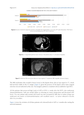
Evolution of patients diagnosed with HCC at the end of treatment
Figure 5. Evolution of patients diagnosed with HCC at the EOT. HCC: hepatocellular carcinoma; AFP: alpha-fetoprotein; TACE: trans-
arterial chemoembolization; EOT: end of treatment
Figure 6. CT image showing three nodular lesions, in the arterial phase. CT: computed tomography
Figure 7. Abdominal MRI showing liver lesions in hypo-signal on T1 sequences. MRI: magnetic resonance imaging
The MRI exploration also revealed a lesion located in the hepatic dome, also in hypo-signal on T1, which
had not been visible on the CT images [Figure 8]; given the presence of the hepatic dome mass, surgical
resection was not indicated in this case. This imagistic pattern is consistent with an infiltrative type HCC.
All the patients that presented high levels of AFP at SVR (12 weeks after the EOT), also underwent
chemoembolization, with one month follow-up showing no tumor progression and decreased AFP
levels. The one patient with normal AFP levels was also evaluated by MRI due to iodine allergy and
underwent one session of chemoembolization, with excellent results - no tumor rebound 9 months
after the procedure.
Figure 9 shows the evolution of all three patients with elevated levels of AFP at 3 months after ending the
antiviral treatment.