
Page 36 - Read Online
P. 36
Iliescu et al. Hepatoma Res 2018;4:3 I http://dx.doi.org/10.20517/2394-5079.2017.48 Page 5 of 10
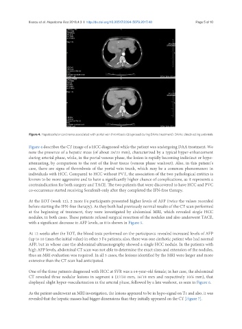
Figure 4. Hepatocellular carcinoma associated with portal vein thrombosis (diagnosed during DAAs treatment). DAAs: direct-acting antivirals
Figure 4 describes the CT image of a HCC diagnosed while the patient was undergoing DAA treatment. We
note the presence of a hepatic mass (of about 28/23 mm), characterized by a typical hyper-enhancement
during arterial phase, while, in the portal venous phase, the lesion is rapidly becoming indistinct or hypo-
attenuating, by comparison to the rest of the liver tissue (venous phase washout). Also, in this patient’s
case, there are signs of thrombosis of the portal vein trunk, which may be a common phenomenon in
individuals with HCC. Compared to HCC without PVT, the association of the two pathological entities is
known to be more aggressive and to have a significantly higher chance of complications, as it represents a
contraindication for both surgery and TACE. The two patients that were discovered to have HCC and PVC
co-occurrence started receiving Sorafenib only after they completed the IFN-free therapy.
At the EOT (week 12), 2 more F4 participants presented higher levels of AFP (twice the values recorded
before starting the IFN-free therapy). As they both had previously normal results of the CT scan performed
at the beginning of treatment, they were investigated by abdominal MRI, which revealed single HCC
nodules, in both cases. These patients refused surgical resection of the nodules and also underwent TACE,
with a significant decrease in AFP levels, as it is shown in Figure 5.
At 12 weeks after the EOT, the blood tests performed on the participants revealed increased levels of AFP
(up to 10 times the initial value) in other 3 F4 patients; also, there was one cirrhotic patient who had normal
AFP, but in whose case the abdominal ultrasonography showed a single HCC nodule. In the patients with
high AFP levels, abdominal CT scan was not able to determine the exact sizes and extension of the nodules,
thus an MRI evaluation was required. In all 3 cases, the lesions identified by the MRI were larger and more
extensive than the CT scan had anticipated.
One of the three patients diagnosed with HCC at SVR was a 64-year-old female; in her case, the abdominal
CT revealed three nodular lesions in segment 8 (27/30 mm, 16/18 mm and respectively 10/6 mm), that
displayed slight hyper-vascularization in the arterial phase, followed by a late washout, as seen in Figure 6.
As the patient underwent an MRI investigation, the lesions appeared to be in hypo-signal on T1 and also, it was
revealed that the hepatic masses had bigger dimensions than they initially appeared on the CT [Figure 7].