
Page 73 - Read Online
P. 73
interleukin-6 in hepatic stem/progenitor cells can cause Notch-1 signaling increases the death receptor 5 (DR5)
[24]
HCC. TGF-β inhibits cell proliferation and promotes expression with augmentation of tumor necrosis factor
tumor cell invasion. Many studies have reported a (TNF)-related apoptosis-inducing ligand induced apoptosis
reduction of TGF-β receptors in up to 70% of HCCs that in vitro and in vivo. [27]
also correlated with metastasis within the liver. On
the other hand, high TGF-β levels have been correlated Sonic Hedgehog pathway
with advanced clinical stages of HCC. This twofold role Activation of Hedgehog signalling is related to liver
[28]
of TGF-β signaling in HCC is explained by the tumor cancer. Up to 60% of human HCCs express Sonic
microenvironment and selective loss of TGF-β-induced Hedgehog. After specific blockade of the sonic Hedgehog
antiproliferative pathway. Tumor cells that have selectively pathway, concomitant down regulation of Gli-related target
lost their growth-inhibitory response to TGF-β, but genes is observed. Furthermore, tumorigenic activation of
retain a functional TGF-β signaling pathway may exhibit SMO can mediate over expression of c-myc, a gene having
increased migration and invasive behaviour on TGF-β an important pathogenic role in liver carcinogenesis.
stimulation. Cells with dysfunctional TGF-β signaling have
been reported to be cancer progenitor cells giving rise to miRNAs
HCC. [25] miRNAs directly interact with specific messenger
RNAs (mRNAs) through base pairing and inhibiting
The Notch signaling pathway the expression of target genes. MiRNAs can undergo
This plays an important role in stem cell self-renewal anomalous regulation during carcinogenesis, and can act
and differentiation. Notch signaling is important in liver as oncogenes or tumor suppressor genes. MiR-181 also
embryogenesis, bile duct formation; angiogenesis and regulates the Wnt/β-catenin signaling pathway with a
endothelial sprouting. However, other signaling pathways positive feedback loop within stem cells. This is used by
have a control on whether Notch functions as a tumor cancer cells to self-propagate continuously, metastasize
suppressor or oncogene. The increased expression of and develop drug resistance.
[26]
genes involved in this pathway has been shown in CD133-
positive liver cancer cells vs. CD133-negative cells. The HEPATOBLASTOMA
activated intracellular form of Notch-3, and the Notch
ligand Jagged, is highly expressed in HCC. Activation of The best characterized pathways in pathogenesis of HB
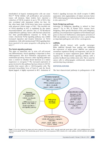
Figure 1: Flow chart depicting the various anti-cancer properties of curcumin. VEGF: vascular endothelial growth factor; MMP: matrix metalloproteinase; PDGF:
platelet derived growth factor; TGF: transforming growth factor; COX: cyclooxygenase; iNOS: inducible nitric oxide synthase; EpCAM: epithelial cell adhesion
molecule
64 Hepatoma Research | Volume 2 | March 9, 2016