
Page 95 - Read Online
P. 95
was placed on the right side. More trocars were used if it was After finishing the nodule ablation, the intrahepatic needle track
necessary to mobilize the liver, perform adhesiolysis between was treated with thermocoagulation to avoid track seeding.
the liver and other adjacent organs or release an associated
procedure like cholecystectomy. Follow-up and defi nition of clinical outcome
Treatment outcome was evaluated with an enhanced CT scan
[14]
Intra-operative laparoscopic ultrasound of the entire liver 2 months after laparoscopic RF. The inflammatory reaction
parenchyma was performed to confirm location of the tumor makes the proper assessment of the treated nodule difficult
to be treated and to rule out the presence of new nodules. during the 1st month. Thereafter, patients were followed with
In cases of lesions that were not visible by ultrasound, a CT or magnetic resonance imaging and with the α-fetoprotein
piece of a 1-cm (22G) needle was inserted preoperatively every 3 months during the 1st year and every 6 months in
into the tumor guided by computed tomography (CT) or the 2nd year.
ultrasound with a signal enhancer. We did not perform
an intra-operative biopsy of the tumor prior to the RFA. Initial incomplete ablation was determined as the presence
Interventional radiologists came to the operating room to of enhanced areas within the treated nodule in the first
follow-up imaging. Sustained complete ablation was defined
perform the RFA procedure and the IOLUS.
as the absence of enhanced areas within the treated area at
the end of the follow-up period.
The RFA was carried out using the Cool-Tip RFA system
(Covidien, Boulder, CO, USA), which uses internally cooled Local tumor recurrence was defined as the presence of a
electrodes (ICEs) for ablation. This system circulates chilled growing tumor in the ablation zone after complete ablation
saline to the tip of the needle electrode, thus lowering had been determined in the first follow-up CT. Distant
the temperature of the tissue immediately adjacent to it, recurrence or new tumor progression was determined as
minimizing tissue charring, and improving the delivery of a growing nodule occurring away from the ablation zone.
energy to surrounding tissues. [13]
Statistical analysis
The RFA electrode was inserted through a separate Data for all patients with HCC were recorded prospectively
percutaneous puncture and the needle was placed as parallel and introduced into a Microsoft ACCESS database.
as possible to the plane of the ultrasound so that its entire Data from patients undergoing laparoscopic RFA were
path could be seen on the ultrasound image as it traversed analyzed retrospectively using the statistical software
the liver parenchyma. A single needle 20-cm long and with SPSS version 18 (SPSS Inc., Chicago, IL, USA). Continuous
a 2-cm tip exposure was used for tumors < 3 cm, and for data were described as means and analyzed with Student’s
tumors ≥ 3 cm a cluster was used to achieve an adequate t-test if the distribution was normal or with Mann-Whitney
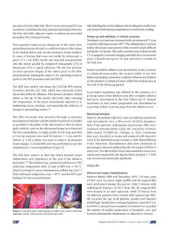
tissue margin. A 12-min RFA cycle was performed as per the U-test otherwise. Discontinuous data were presented as
manufacturer’s recommendations [Figure 2]. percentages and were analyzed by the Chi-square of Fisher’s
exact test. Overall survival curves and cumulative recurrence
The ICEs have sensors in their tips which measure tissue curves were analyzed by the Kaplan-Meier method. P < 0.05
temperature and impedance at the end of the ablation was considered statistically significant.
[13]
procedure. The ablation was considered satisfactory if the
end-tissue temperature after 12 min of RFA was ≥ 60 °C, RESULTS
which is enough to cause instantaneous cellular necrosis.
[13]
If the end-tissue temperature was < 60 °C, another RFA cycle Patient and tumor characteristics
lasting 6-10 min was performed. Between March 2009 and December 2014, 149 new cases
of HCC were recorded. Eight perRFA and 40 surgical RFA
were performed during this period. Only 3 lesions with
radiological features of HCC from the 40 surgical RFA
were treated by an open approach, while 37 lesions from
32 different patients were treated with laparoscopic RFA.
We recorded the age of all patients, gender, liver function
(Child-Pugh Classification), etiology [hepatitis C virus (HCV ) or
+
+
a b non-HCV ], and previous treatment. For tumors we recorded
Figure 2: Surgical images of radiofrequency ablation of (a) a lesion next to the the number of nodules (uninodular or binodular), size, and
diaphragm and (b) a lesion close to duodenum and colon location (subcapsular, intrahepatic or adjacent to viscera).
88 Hepatoma Research | Volume 1 | Issue 2 | July 15, 2015