
Page 39 - Read Online
P. 39
®
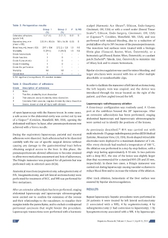
Table 2: Perioperative results scalpel (Harmonic Ace Shears ; Ethicon, Endo-Surgery,
Group 1 Group 2 P S/NS Cincinnati, OH, USA) or with a vessel sealer (Enseal Tissue
®
(%) (%) Sealer ; Ethicon, Endo-Surgery, Cincinnati, OH, USA)
Extensive adhesions 5 (71.4) 2 (11.7) 0.01 S or (Ligasure™; Covidien, Mansfield, MA, USA), and was
(grade 3-4)
Operative time, min 220.14 ± 80.06 150 ± 56.18 0.03 S performed with reduced bleeding, due to a reduction of
(mean ± SD) portal inflow of up to 30% because of the pneumoperitoneum.
Blood loss, mL (mean ± SD) 297 ± 134 272.2 ± 120 1.0 NS The resection bed surfaces were treated with a biologic
Morbidity 5 (29.4) 2 (28.5) 1.0 NS fibrin glue (Tissucol; Baxter, Wien, Österreich), or a
Grade I atelectasis 1 1 - -
hemostatic gel (Floseal; Baxter, Wien, Österreich), or a sealant
Grade I ascites 1 0 - - patch (TachoSil ; Takeda, Linz, Österreich) to minimize risk
®
Grade II pneumonia 2 0 - -
of biliary leak and to ensure hemostasis.
Grade II bleeding 1 0 - -
Grade IIIa perforation 0 1 - -
Mortality Nil Nil - - Bipolar electrocoagulation was used for minor bleeding, and
Conversion 1 0 - - larger structures were secured with ties or either multiple
S/NS: signifi cant/nonsignifi cant; SD: standard deviation absorbable or nonabsorbable clips.
Table 3: Classifi cation of adhesions In order to facilitate the maneuver of left lateral sectionectomy,
Grade Description of adhesions the left hepatic vein was stapled, and the device was
0 None
introduced through the trocar located on the right of the
1 Thin fi lm, divided by blunt dissection
patient, and then angled toward the left.
2 Thin vascular, easily divided by sharp dissection
3 Extensive thick vascular, requires division by sharp dissection
Laparoscopic radiofrequency ablation
4 Dense, bowel at risk of injury with division
A three-trocar configuration was routinely used. A 12-mm
port at the umbilicus housed the 30° laparoscope. After
of open laparoscopy with the Hasson trocar. In some cases,
a safe access to the abdominal cavity was carried out by use an extensive adhesiolysis has been performed, staging
of a Visiport (Covidien, Mansfield, MA, USA), opening the abdominal laparoscopy and laparoscopic ultrasonography
®
abdominal wall layer by layer, after pneumoperitoneum was were carried out to identify the positions of the lesions.
achieved with a Verres needle.
[9]
As previously described, RFA was carried out with
During the exploratory laparoscopy, parietal and visceral multi-electrode 15-gauge radiofrequency probes (RITA Medical
adhesions were dissected. Such adhesions had to be dissected Systems, Mountain View, CA, USA). Hook-shaped retractable
carefully with the use of specific surgical devices without electrodes were deployed to a maximum diameter of 3 cm.
causing any damage to the gastrointestinal tract before After every electrode had reached a temperature of 100 °C,
obtaining surgical access to the liver. In this phase, the the ablation was performed in a step-by-step fashion, with a
pneumoperitoneum allowed adhesions to become strained single step lasting approximately 8-10 min. In two patients
to allow more meticulous assessment and lysis of adherences. with a deep HCC, the size of the lesion was slightly larger
The Pringle maneuver was prepared for all patients but was than that recommended for a standard RFA (35 and 33 mm,
performed only in selected cases (8/24). respectively). In these two cases, a Pringle maneuver was
carried out during laparoscopy causing vascular occlusion to
Anatomical resections (segmentectomy, subsegmentctomy of reduce blood flow and to increase the volume of the ablation.
IVb, bisegmentectomy, and left lateral sectionectomy) were
performed for treatment of HCC, and WR was performed for After track ablation, hemostasis of the liver surface was
liver metastases. ensured by bipolar electrocoagulation.
After an extensive adhesiolysis has been performed, staging RESULTS
abdominal laparoscopy and laparoscopic ultrasonography
were carried out to confirm the extension of the lesions Repeat laparoscopic hepatic procedures were performed in
and their relationships to the vasculature, to visualize their 24 patients: 6 were treated by left lateral sectionectomy
margins inside the parenchyma, and to exclude a widespread (1 associated with a WR), 4 by segmentectomy, 4 by
peritoneal carcinosis that might hinder the procedure. subsegmentectomy (1 had conversion to laparotomy), 1 by
Laparoscopic transections were performed with a harmonic bysegmentectomy associated with a WR, 4 by laparoscopic
32 Hepatoma Research | Volume 1 | Issue 1 | April 15, 2015