
Page 99 - Read Online
P. 99
Page 4 of 16 Cadamuro et al. Hepatoma Res 2022;8:11 https://dx.doi.org/10.20517/2394-5079.2021.140
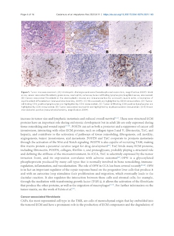
Figure 1. Tumor microenvironment. (A) Intrahepatic cholangiocarcinoma (hematoxylin and eosin stain, magnification 200×): black
arrow, cancer-associated fibroblasts; green arrow, neutrophils; red arrow, tumor-infiltrating lymphocytes; long black arrow, microvessel.
(B) Cancer-associated fibroblasts in the desmoplastic stroma are immunoreactive for α-smooth muscle actin, a biomarker of
myofibroblast differentiation (immunohistochemistry, 200×). (C) Microvessels are highlighted by CD34 immunostain. (D) Tumor-
infiltrating CD4 positive lymphocytes are highlighted by CD4 immunostain. (E) Tumor-infiltrating CD8 positive lymphocytes are
highlighted by CD8 immunostain. (F) Tumor-associated neutrophils are highlighted by myeloperoxidase immunostain. (B-F) Brown
stain indicates positive (mmunohistochemistry, magnification 200×).
increase in tumor size and lymphatic metastasis and reduced overall survival [3,12] . These non-structural ECM
proteins have an important role during embryonic development but in adult life are only expressed during
tissue remodeling and wound repair [18-20] . POSTN can act as both a promoter and a suppressor of cancer cell
invasiveness, interacting with other ECM proteins, such as collagen types I and V, fibronectin, TnC, and
heparin, and contribute to the activation of pathways of tissue remodeling, fibrogenesis, cell motility,
angiogenesis, tumor invasiveness, and metastasis. POSTN and TnC cooperate to promote metastasis
through the activation of the Wnt and Notch signaling. POSTN is also capable of recruiting TAM, making
[21]
this matrix protein a potential curative target for drug development . TnC binds many ECM proteins,
including fibronectin, POSTN, collagen, fibrillin-2, and proteoglycans, probably playing a structural role
and defining the stiffness of the microenvironment. In iCCA, TnC is selectively expressed by the tumor
invasion front, and its expression correlates with adverse outcomes . OPN is a glycosylated
[22]
phosphoprotein produced by many cell types that is normally involved in bone remodeling, immune-
regulation, inflammation, and vascularization. The role of OPN in CCA has been covered recently [23,24] . OPN
is in fact an important regulator of the repair response based on the progenitor liver cells that produce it
and with an autocrine loop stimulates their proliferation and migration, which eventually leads to the
ductular reaction. It also regulates the interaction between these cells and stromal cells; for example,
through the mediation with transforming growth factor (TGF) β, it allows the activation of the fibroblasts
that produce the other proteins, as well as the migration of macrophages [23,24] . For further information on the
tumor matrix, see the work of Fabris et al. .
[25]
Cancer-associated fibroblasts
CAFs, the most represented cell type in the TME, are cells of mesenchymal origin that lay embedded into
the tumoral ECM and have a prominent role in the production of ECM components and the degradation of