
Page 98 - Read Online
P. 98
Cadamuro et al. Hepatoma Res 2022;8:11 https://dx.doi.org/10.20517/2394-5079.2021.140 Page 3 of 16
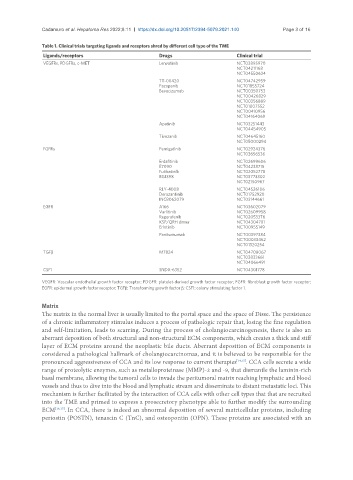
Table 1. Clinical trials targeting ligands and receptors shred by different cell type of the TME
Ligands/receptors Drugs Clinical trial
VEGFRs, PDGFRs, c-MET Lenvatinib NCT03895970
NCT04211168
NCT04550624
TT-00420 NCT04742959
Pazopanib NCT01855724
Bevacizumab NCT00350753
NCT00426829
NCT00356889
NCT01007552
NCT00410956
NCT04164069
Apatinib NCT03251443
NCT04454905
Tivozanib NCT04645160
NCT05000294
FGFRs Pemigatinib NCT02924376
NCT03656536
Erdafitinib NCT02699606
E7090 NCT04238715
Futibatinib NCT02052778
BGJ398 NCT03773302
NCT02150967
RLY-4008 NCT04526106
Derazantinib NCT01752920
INCB062079 NCT03144661
EGFR A166 NCT03602079
Varlitinib NCT02609958
Regorafenib NCT02053376
KSP/QRH dimer NCT04304781
Erlotinib NCT00955149
Panitumumab NCT00397384
NCT00033462
NCT01320254
TGFβ M7824 NCT04708067
NCT03833661
NCT04066491
CSF1 SNDX-6352 NCT04301778
VEGFR: Vascular endothelial growth factor receptor; PDGFR: platelet-derived growth factor receptor; FGFR: fibroblast growth factor receptor;
EGFR: epidermal growth factor receptor; TGFβ: Transforming growth factor β; CSF1: colony stimulating factor 1.
Matrix
The matrix in the normal liver is usually limited to the portal space and the space of Disse. The persistence
of a chronic inflammatory stimulus induces a process of pathologic repair that, losing the fine regulation
and self-limitation, leads to scarring. During the process of cholangiocarcinogenesis, there is also an
aberrant deposition of both structural and non-structural ECM components, which creates a thick and stiff
layer of ECM proteins around the neoplastic bile ducts. Aberrant deposition of ECM components is
considered a pathological hallmark of cholangiocarcinomas, and it is believed to be responsible for the
pronounced aggressiveness of CCA and its low response to current therapies [14,15] . CCA cells secrete a wide
range of proteolytic enzymes, such as metalloproteinase (MMP)-2 and -9, that dismantle the laminin-rich
basal membrane, allowing the tumoral cells to invade the peritumoral matrix reaching lymphatic and blood
vessels and thus to dive into the blood and lymphatic stream and disseminate to distant metastatic loci. This
mechanism is further facilitated by the interaction of CCA cells with other cell types that that are recruited
into the TME and primed to express a prosecretory phenotype able to further modify the surrounding
ECM [16,17] . In CCA, there is indeed an abnormal deposition of several matricellular proteins, including
periostin (POSTN), tenascin C (TnC), and osteopontin (OPN). These proteins are associated with an