
Page 84 - Read Online
P. 84
Page 4 of 15 Della Corte et al. Hepatoma Res 2022;8:5 https://dx.doi.org/10.20517/2394-5079.2021.103
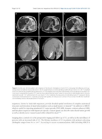
Figure 1. Seventy-year-old male patient with diagnosis of intrahepatic cholangiocarcinoma (ICC) undergoing chemotherapy and drug-
eluting beads transarterial chemoembolization. (A) T2 weighted axial scan demonstrating multifocal hyperintense ICC; (B) T1 weighted
axial scan demonstrating multifocal hypointense ICC; (C) T2 weighted axial scan after chemotherapy showing significant reduction; (D)
cone beam computed tomography (CT) arterial phase demonstrating target lesion for transarterial chemoembolization showing
peripheral vascular “rim” enhancement; (E) cone beam CT after treatment showing uptake of embolizing beads; (F) angiogram during
transarterial chemoembolization treatment, showing lesion uptake; (G) delayed post-contrast CT scan phase 1 month after treatment
demonstrating complete devascularization of the lesion.
sequences, driven by thick slab sequences, provide detailed spatial resolution of complex anatomical
structures and detection of small abnormalities such as small masses or stenosis . In addition to MRCP,
[33]
which is useful for depicting intraductal CC tumor growth, DWI-MRI, dynamic contrast-enhanced MRI,
and late-phase sequences with hepatocyte-specific contrast agents can describe extra-ductal tumor growth
as well as identify tumor masses located within dilated bile ducts [33,34] .
Imaging plays a central role in the preoperative staging and follow-up of CC, as well as in the surveillance of
patients with an increased risk of CC. The lifetime incidence of CC in patients with primary sclerosing
[33]
cholangitis ranges from 5% to 10% . According to recent recommendations, MRI including MRCP is